|
Comment citer 1. Pour la citation dans le texte (Matériel & Méthodes) : 2. Pour le tableau des ressources clés : |
||
|
Numéro vert : (877) 796-6397 -- États-Unis et Canada uniquement -- |
Fax : +1-832-582-8590 Commandes : +1-832-582-8158 |
Support technique : +1-832-582-8158 Ext:3 Veuillez indiquer votre numéro de commande dans le-mail. Nous nous efforçons de répondre à toutes les demandes par e-mail dans un délai dun jour ouvrable. |
Description biologique
| Spécificité | HACE1 Antibody [A17B18] reconnaît les niveaux endogènes de la protéine HACE1 totale. |
|---|---|
| Contexte | Le gène HACE1 (HECT domain and ankyrin repeat-containing E3 ubiquitin-protein ligase 1) est situé sur le chromosome 6 et code pour une protéine composée de 909 acides aminés. HACE1 est fortement exprimé dans divers tissus humains, y compris le cœur, le cerveau, le placenta, le pancréas et les reins fœtaux et adultes. L'ARNm de HACE1 est exprimé de manière ubiquitaire dans les tissus humains normaux. Des niveaux d'expression réduits de HACE1 sont fortement associés à l'hyperméthylation de deux îlots CpG situés en amont de son locus génique, suggérant qu'une régulation épigénétique joue un rôle dans son silençage. HACE1 est fréquemment régulé à la baisse dans plusieurs types de cancer, y compris le lymphome à cellules T/natural killer (NKTCL), le cancer colorectal et le cancer gastrique. Au niveau subcellulaire, HACE1 se localise principalement dans le réticulum endoplasmique et le cytosol, bien que seuls de faibles niveaux de protéine endogène soient généralement détectés. Fonctionnellement, HACE1 agit comme une E3 Ligase et s'associe à l'enzyme E2 UBCH7 pour catalyser l'ubiquitination des protéines cibles. Il est notamment impliqué dans la dégradation dépendante de la phosphorylation de la cycline D1, jouant ainsi un rôle dans l'inhibition de la progression du cycle cellulaire. HACE1 se lie également préférentiellement à la forme active, liée au GTP, de la petite GTPase Rac1 et favorise sa polyubiquitination. Cette activité est essentielle pour la dégradation de Rac1 en réponse au facteur nécrosant cytotoxique 1 (CNF1), facilitant l'invasion bactérienne des monocouches de cellules endothéliales, et soulignant un rôle pour HACE1 dans les mécanismes de défense immunitaire innée. La perte de HACE1 conduit au développement spontané de tumeurs à apparition tardive, soutenant davantage sa fonction de suppresseur de tumeur. Le gène se trouve dans la région chromosomique 6q21 qui est un point chaud impliqué dans plusieurs cancers humains, soulignant son importance clinique dans la tumorigenèse. |
Informations dutilisation
| Application | WB | Dilution |
|
||
|---|---|---|---|---|---|
| Réactivité | Mouse, Rat, Human | ||||
| Source | Rabbit Monoclonal Antibody | MW | 102 kDa | ||
| Tampon de stockage | PBS, pH 7.2+50% Glycerol+0.05% BSA+0.01% NaN3 | Stockage (À partir de la date de réception) |
-20°C (avoid freeze-thaw cycles), 2 years | ||
| WB |
Experimental Protocol:
Sample preparation
1. Tissue: Lyse the tissue sample by adding an appropriate volume of ice-cold RIPA/NP-40 Lysis Buffer (containing Protease Inhibitor Cocktail),and homogenize the tissue at a low temperature. 2. Adherent cell: Aspirate the culture medium and wash the cells with ice-cold PBS twice. Lyse the cells by adding an appropriate volume of RIPA/NP-40 Lysis Buffer (containing Protease Inhibitor Cocktail) and put the sample on ice for 5 min. 3. Suspension cell: Transfer the culture medium to a pre-cooled centrifuge tube. Centrifuge and aspirate the supernatant. Wash the cells with ice-cold PBS twice. Lyse the cells by adding an appropriate volume of RIPA/NP-40 Lysis Buffer (containing Protease Inhibitor Cocktail) and put the sample on ice for 5 min. 4. Place the lysate into a pre-cooled microcentrifuge tube. Centrifuge at 4°C for 15 min. Collect the supernatant;
5. Remove a small volume of lysate to determine the protein concentration;
6. Combine the lysate with protein loading buffer. Boil 20 µL sample under 95-100°C for 5 min. Centrifuge for 5 min after cool down on ice.
Electrophoretic separation
1. According to the concentration of extracted protein, load appropriate amount of protein sample and marker onto SDS-PAGE gels for electrophoresis. Recommended separating gel (lower gel) concentration: 5%. Reference Table for Selecting SDS-PAGE Separation Gel Concentrations 2. Power up 80V for 30 minutes. Then the power supply is adjusted (110 V~150 V), the Marker is observed, and the electrophoresis can be stopped when the indicator band of the predyed protein Marker where the protein is located is properly separated. (Note that the current should not be too large when electrophoresis, too large current (more than 150 mA) will cause the temperature to rise, affecting the result of running glue. If high currents cannot be avoided, an ice bath can be used to cool the bath.)
Transfer membrane
1. Take out the converter, soak the clip and consumables in the pre-cooled converter;
2. Activate PVDF membrane with methanol for 1 min and rinse with transfer buffer;
3. Install it in the order of "black edge of clip - sponge - filter paper - filter paper - glue -PVDF membrane - filter paper - filter paper - sponge - white edge of clip"; 4. The protein was electrotransferred to PVDF membrane. ( 0.45 µm PVDF membrane is recommended ) Reference Table for Selecting PVDF Membrane Pore Size Specifications Recommended conditions for wet transfer: 200 mA, 120 min. ( Note that the transfer conditions can be adjusted according to the protein size. For high-molecular-weight proteins, a higher current and longer transfer time are recommended. However, ensure that the transfer tank remains at a low temperature to prevent gel melting.)
Block
1. After electrotransfer, wash the film with TBST at room temperature for 5 minutes;
2. Incubate the film in the blocking solution for 1 hour at room temperature;
3. Wash the film with TBST for 3 times, 5 minutes each time.
Antibody incubation
1. Use 5% skim milk powder to prepare the primary antibody working liquid (recommended dilution ratio for primary antibody 1:1000), gently shake and incubate with the film at 4°C overnight; 2. Wash the film with TBST 3 times, 5 minutes each time;
3. Add the secondary antibody to the blocking solution and incubate with the film gently at room temperature for 1 hour;
4. After incubation, wash the film with TBST 3 times for 5 minutes each time.
Antibody staining
1. Add the prepared ECL luminescent substrate (or select other color developing substrate according to the second antibody) and mix evenly;
2. Incubate with the film for 1 minute, remove excess substrate (keep the film moist), wrap with plastic film, and expose in the imaging system.
|
Références
|
Données dapplication
WB
Validé par Selleck
-
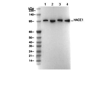 Lane 1: HEK293, Lane 2: SH-SY5Y, Lane 3: Mouse brain, Lane 4: Rat brain