|
Comment citer 1. Pour la citation dans le texte (Matériel & Méthodes) : 2. Pour le tableau des ressources clés : |
||
|
Numéro vert : (877) 796-6397 -- États-Unis et Canada uniquement -- |
Fax : +1-832-582-8590 Commandes : +1-832-582-8158 |
Support technique : +1-832-582-8158 Ext:3 Veuillez indiquer votre numéro de commande dans le-mail. Nous nous efforçons de répondre à toutes les demandes par e-mail dans un délai dun jour ouvrable. |
Description biologique
| Spécificité | Insulin Antibody [K13G15] reconnaît les niveaux endogènes de la protéine Insulin totale. |
|---|---|
| Contexte | L'insuline est une hormone peptidique composée de 51 acides aminés, sécrétée par les cellules β du pancréas. Elle joue un rôle vital dans le maintien de l'homéostasie du glucose en se liant au récepteur de l'insuline (IR), une Protein Tyrosine Kinase transmembranaire composée de deux sous-unités α extracellulaires responsables de la liaison du ligand et de deux sous-unités β intracellulaires qui médient la transduction du signal. Lors de l'engagement de l'insuline, le récepteur subit une autophosphorylation sur ses résidus tyrosine, activant son activité kinase. Cela conduit à la phosphorylation des substrats du récepteur de l'insuline (IRS), qui à leur tour recrutent et activent la phosphoinositide 3-kinase (PI3K). La PI3K activée génère du phosphatidylinositol (3,4,5)-trisphosphate (PIP3), ce qui facilite l'activation d'AKT (protein kinase B). L'AKT activée régule plusieurs processus métaboliques en aval : elle favorise l'absorption du glucose dans les muscles squelettiques et le tissu adipeux en induisant la translocation du transporteur de glucose GLUT4 vers la membrane cellulaire, supprime la production hépatique de glucose en phosphorylant et en inhibant le facteur de transcription FOXO1, et améliore le stockage du glycogène en inhibant la glycogène synthase kinase 3 (GSK3). De plus, la signalisation de l'insuline inhibe la lipolyse par la suppression médiée par PDE3B et ABHD15 de la lipase triglycéride des adipocytes (ATGL) et de la lipase sensible aux hormones (HSL), tout en favorisant simultanément la lipogenèse et la synthèse des protéines via l'activation du complexe mTORC1. La résistance à l'insuline survient lorsque les cellules deviennent moins réactives à la signalisation de l'insuline, souvent en raison d'altérations à plusieurs niveaux de la voie. Celles-ci peuvent inclure une diminution du nombre de récepteurs de l'insuline, une phosphorylation défectueuse de l'IRS ou une signalisation PI3K/AKT perturbée. Les facteurs contributifs comprennent l'inflammation chronique, le stress oxydatif et l'accumulation d'intermédiaires lipidiques tels que les diacylglycérols et les céramides, qui interfèrent avec l'activité d'IRS-1 et d'AKT. En outre, le dépôt lipidique ectopique dans des tissus comme le foie et les muscles squelettiques peut activer des kinases liées au stress, y compris JNK et PKCθ, conduisant à la phosphorylation de la sérine des protéines IRS et à l'inhibition subséquente de la signalisation de l'insuline. Le dysfonctionnement mitochondrial et le stress du réticulum endoplasmique (RE) contribuent également à l'altération du métabolisme du glucose, entraînant une hyperglycémie persistante et un dysfonctionnement progressif des cellules β – des caractéristiques du diabète de type 2. |
Informations dutilisation
| Application | WB, IHC, IF, FCM | Dilution |
|
||||||||
|---|---|---|---|---|---|---|---|---|---|---|---|
| Réactivité | Human, Mouse, Rat | ||||||||||
| Source | Rabbit Monoclonal Antibody | MW | 12 kDa | ||||||||
| Tampon de stockage | PBS, pH 7.2+50% Glycerol+0.05% BSA+0.01% NaN3 | Stockage (À partir de la date de réception) |
-20°C (avoid freeze-thaw cycles), 2 years | ||||||||
| IF |
Experimental Protocol:
Specimen Preparation
1. Aspirate liquid, then cover cells to a depth of 2–3 mm with 4% Paraformaldehyde diluted in 1X PBS.
NOTE: Paraformaldehyde is toxic, use only in a fume hood.
2. Fix cells for 15 min at room temperature.
3. Aspirate fixative, rinse three times in 1X PBS for 5 min each.
4. Proceed with Immunostaining.
Immunostaining
1. Add theblocking buffer and incubate for 60 min at RT.
2. Prepare primary antibody diluent in antibody dilution buffer as recommended .
3. Aspirate blocking solution, apply diluted primary antibody.
4. Incubate overnight at 4°C.
5. Rinse three times in 1X PBS for 5 min each.
6. Incubate specimens in fluorochrome-conjugated secondary antibody diluted in antibody dilution buffer for 1–2 hr at room temperature in the dark.
7. Rinse three times in 1X PBS for 5 min each.
8. Mount slides usingmounting medium with DAPI and cover with coverslips.
9. For best results, allow mountant to cure overnight at room temperature. For long-term storage, store slides flat at 23°C protected from light.
|
| IHC |
Experimental Protocol:
Deparaffinization/Rehydration
1. Deparaffinize/hydrate sections:
2. Incubate sections in three washes of xylene for 5 min each.
3. Incubate sections in two washes of 100% ethanol for 10 min each.
4. Incubate sections in two washes of 95% ethanol for 10 min each.
5. Wash sections two times in dH2O for 5 min each.
6.Antigen retrieval: For Citrate: Heat slides in a microwave submersed in 1X citrate unmasking solution until boiling is initiated; continue with 10 min at a sub-boiling temperature (95°-98°C). Cool slides on bench top for 30 min.
Staining
1. Wash sections in dH2O three times for 5 min each.
2. Incubate sections in 3% hydrogen peroxide for 10 min.
3. Wash sections in dH2O two times for 5 min each.
4. Wash sections in wash buffer for 5 min.
5. Block each section with 100–400 µl of blocking solution for 1 hr at room temperature.
6. Remove blocking solution and add 100–400 µl primary antibody diluent in to each section. Incubate overnight at 4°C.
7. Remove antibody solution and wash sections with wash buffer three times for 5 min each.
8. Cover section with 1–3 drops HRPas needed. Incubate in a humidified chamber for 30 min at room temperature.
9. Wash sections three times with wash buffer for 5 min each.
10. Add DAB Chromogen Concentrate to DAB Diluent and mix well before use.
11. Apply 100–400 µl DAB to each section and monitor closely. 1–10 min generally provides an acceptable staining intensity.
12. Immerse slides in dH2O.
13. If desired, counterstain sections with hematoxylin.
14. Wash sections in dH2O two times for 5 min each.
15. Dehydrate sections: Incubate sections in 95% ethanol two times for 10 sec each; Repeat in 100% ethanol, incubating sections two times for 10 sec each; Repeat in xylene, incubating sections two times for 10 sec each.
16. Mount sections with coverslips and mounting medium.
|
| IF |
Experimental Protocol:
Sample Preparation
1. Adherent Cells: Place a clean, sterile coverslip in a culture dish. Once the cells grow to near confluence as a monolayer, remove the coverslip for further use.
2. Suspension Cells: Seed the cells onto a clean, sterile slide coated with poly-L-lysine.
3. Frozen Sections: Allow the slide to thaw at room temperature. Wash it with pure water or PBS for 2 times, 3 minutes each time.
4. Paraffin Sections: Deparaffinization and rehydration. Wash the slide with pure water or PBS for 3 times, 3 minutes each time. Then perform antigen retrieval.
Fixation
1. Fix the cell coverslips/spots or tissue sections at room temperature using a fixative such as 4% paraformaldehyde (4% PFA) for 10-15 minutes.
2. Wash the sample with PBS for 3 times, 3 minutes each time.
Permeabilization
1.Add a detergent such as 0.1–0.3% Triton X-100 to the sample and incubate at room temperature for 10–20 minutes.
(Note: This step is only required for intracellular antigens. For antigens expressed on the cell membrane, this step is unnecessary.)
Wash the sample with PBS for 3 times, 3 minutes each time.
Blocking
Add blocking solution and incubate at room temperature for at least 1 hour. (Common blocking solutions include: serum from the same source as the secondary antibody, BSA, or goat serum.)
Note: Ensure the sample remains moist during and after the blocking step to prevent drying, which can lead to high background.
Immunofluorescence Staining (Day 1)
1. Remove the blocking solution and add the diluted primary antibody.
2. Incubate the sample in a humidified chamber at 4°C overnight.
Immunofluorescence Staining (Day 2)
1. Remove the primary antibody and wash with PBST for 3 times, 5 minutes each time.
2. Add the diluted fluorescent secondary antibody and incubate in the dark at 4°C for 1–2 hours.
3. Remove the secondary antibody and wash with PBST for 3 times, 5 minutes each time.
4. Add diluted DAPI and incubate at room temperature in the dark for 5–10 minutes.
5. Wash with PBST for 3 times, 5 minutes each time.
Mounting
1. Mount the sample with an anti-fade mounting medium.
2. Allow the slide to dry at room temperature overnight in the dark.
3. Store the slide in a slide storage box at 4°C, protected from light.
|
Références
|
Données dapplication
WB
Validé par Selleck
-
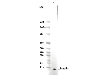 Lane 1: INS-1