|
Hoe te citeren 1. Voor in-tekst citatie (materialen en methoden): 2. Voor de tabel met belangrijke bronnen: |
||
|
Gratis nummer: (877) 796-6397 -- Alleen VS en Canada -- |
Fax: +1-832-582-8590 Bestellingen: +1-832-582-8158 |
Technische ondersteuning: +1-832-582-8158 Ext:3 Gelieve uw bestelnummer in de e-mail te vermelden. Wij streven ernaar alle e-mailvragen binnen één werkdag te beantwoorden. |
Biologische Beschrijving
| Specificiteit | Insulin Antibody [K13G15] herkent endogene niveaus van totaal Insulin-eiwit. |
|---|---|
| Achtergrond | Insuline is een peptidehormoon, samengesteld uit 51 aminozuren, afgescheiden door de β-cellen van de alvleesklier. Het speelt een vitale rol bij het handhaven van glucosehomeostase door te binden aan de insulinereceptor (IR), een transmembraan Protein Tyrosine Kinase die is samengesteld uit twee extracellulaire α-subeenheden die verantwoordelijk zijn voor ligandbinding en twee intracellulaire β-subeenheden die signaaltransductie mediëren. Bij insulinebinding ondergaat de receptor autofosforylering op zijn tyrosineresiduen, waardoor zijn kinase-activiteit wordt geactiveerd. Dit leidt tot fosforylering van insulinereceptorsubstraten (IRS), die op hun beurt fosfoinositide 3-kinase (PI3K) rekruteren en activeren. Geactiveerde PI3K genereert fosfatidylinositol (3,4,5)-trifosfaat (PIP3), wat de activering van AKT (protein kinase B) vergemakkelijkt. Geactiveerd AKT reguleert verschillende stroomafwaartse metabolische processen: het bevordert de glucoseopname in skeletspieren en vetweefsel door de translocatie van de GLUT4-glucosetransporter naar het celmembraan te induceren, onderdrukt de hepatische glucoseproductie door de transcriptiefactor FOXO1 te fosforyleren en te remmen, en verbetert de glycogeenopslag door glycogeen synthase kinase 3 (GSK3) te remmen. Bovendien remt insulinesignalering lipolyse via PDE3B- en ABHD15-gemedieerde onderdrukking van adipocytentriglyceridelipase (ATGL) en hormoongevoelige lipase (HSL), terwijl het tegelijkertijd lipogenese en eiwitsynthese bevordert via activering van het mTORC1-complex. Insulineresistentie treedt op wanneer cellen minder reageren op insulinesignalering, vaak als gevolg van stoornissen op meerdere niveaus van de route. Deze kunnen onder meer een verminderd aantal insulinereceptoren, defecte IRS-fosforylering of verstoorde PI3K/AKT-signalering omvatten. Bijdragende factoren zijn chronische ontsteking, oxidatieve stress en accumulatie van lipide-intermediairen zoals diacylglycerolen en ceramiden, die de IRS-1- en AKT-activiteit verstoren. Bovendien kan ectopische lipide-afzetting in weefsels zoals de lever en skeletspieren stressgerelateerde kinasen, waaronder JNK en PKCθ, activeren, wat leidt tot serinefosforylering van IRS-eiwitten en daaropvolgende remming van insulinesignalering. Mitochondriale disfunctie en endoplasmatisch reticulum (ER)-stress dragen ook bij aan een verminderde glucosemetabolisme, wat leidt tot aanhoudende hyperglykemie en progressieve β-celdisfunctie – kenmerken van type 2 diabetes. |
Gebruiksinformatie
| Toepassing | WB, IHC, IF, FCM | Verdunning |
|
||||||||
|---|---|---|---|---|---|---|---|---|---|---|---|
| Reactiviteit | Human, Mouse, Rat | ||||||||||
| Bron | Rabbit Monoclonal Antibody | MW | 12 kDa | ||||||||
| Opslagbuffer | PBS, pH 7.2+50% Glycerol+0.05% BSA+0.01% NaN3 | Opslag (Vanaf de datum van ontvangst) |
-20°C (avoid freeze-thaw cycles), 2 years | ||||||||
| IF |
Experimental Protocol:
Specimen Preparation
1. Aspirate liquid, then cover cells to a depth of 2–3 mm with 4% Paraformaldehyde diluted in 1X PBS.
NOTE: Paraformaldehyde is toxic, use only in a fume hood.
2. Fix cells for 15 min at room temperature.
3. Aspirate fixative, rinse three times in 1X PBS for 5 min each.
4. Proceed with Immunostaining.
Immunostaining
1. Add theblocking buffer and incubate for 60 min at RT.
2. Prepare primary antibody diluent in antibody dilution buffer as recommended .
3. Aspirate blocking solution, apply diluted primary antibody.
4. Incubate overnight at 4°C.
5. Rinse three times in 1X PBS for 5 min each.
6. Incubate specimens in fluorochrome-conjugated secondary antibody diluted in antibody dilution buffer for 1–2 hr at room temperature in the dark.
7. Rinse three times in 1X PBS for 5 min each.
8. Mount slides usingmounting medium with DAPI and cover with coverslips.
9. For best results, allow mountant to cure overnight at room temperature. For long-term storage, store slides flat at 23°C protected from light.
|
| IHC |
Experimental Protocol:
Deparaffinization/Rehydration
1. Deparaffinize/hydrate sections:
2. Incubate sections in three washes of xylene for 5 min each.
3. Incubate sections in two washes of 100% ethanol for 10 min each.
4. Incubate sections in two washes of 95% ethanol for 10 min each.
5. Wash sections two times in dH2O for 5 min each.
6.Antigen retrieval: For Citrate: Heat slides in a microwave submersed in 1X citrate unmasking solution until boiling is initiated; continue with 10 min at a sub-boiling temperature (95°-98°C). Cool slides on bench top for 30 min.
Staining
1. Wash sections in dH2O three times for 5 min each.
2. Incubate sections in 3% hydrogen peroxide for 10 min.
3. Wash sections in dH2O two times for 5 min each.
4. Wash sections in wash buffer for 5 min.
5. Block each section with 100–400 µl of blocking solution for 1 hr at room temperature.
6. Remove blocking solution and add 100–400 µl primary antibody diluent in to each section. Incubate overnight at 4°C.
7. Remove antibody solution and wash sections with wash buffer three times for 5 min each.
8. Cover section with 1–3 drops HRPas needed. Incubate in a humidified chamber for 30 min at room temperature.
9. Wash sections three times with wash buffer for 5 min each.
10. Add DAB Chromogen Concentrate to DAB Diluent and mix well before use.
11. Apply 100–400 µl DAB to each section and monitor closely. 1–10 min generally provides an acceptable staining intensity.
12. Immerse slides in dH2O.
13. If desired, counterstain sections with hematoxylin.
14. Wash sections in dH2O two times for 5 min each.
15. Dehydrate sections: Incubate sections in 95% ethanol two times for 10 sec each; Repeat in 100% ethanol, incubating sections two times for 10 sec each; Repeat in xylene, incubating sections two times for 10 sec each.
16. Mount sections with coverslips and mounting medium.
|
| IF |
Experimental Protocol:
Sample Preparation
1. Adherent Cells: Place a clean, sterile coverslip in a culture dish. Once the cells grow to near confluence as a monolayer, remove the coverslip for further use.
2. Suspension Cells: Seed the cells onto a clean, sterile slide coated with poly-L-lysine.
3. Frozen Sections: Allow the slide to thaw at room temperature. Wash it with pure water or PBS for 2 times, 3 minutes each time.
4. Paraffin Sections: Deparaffinization and rehydration. Wash the slide with pure water or PBS for 3 times, 3 minutes each time. Then perform antigen retrieval.
Fixation
1. Fix the cell coverslips/spots or tissue sections at room temperature using a fixative such as 4% paraformaldehyde (4% PFA) for 10-15 minutes.
2. Wash the sample with PBS for 3 times, 3 minutes each time.
Permeabilization
1.Add a detergent such as 0.1–0.3% Triton X-100 to the sample and incubate at room temperature for 10–20 minutes.
(Note: This step is only required for intracellular antigens. For antigens expressed on the cell membrane, this step is unnecessary.)
Wash the sample with PBS for 3 times, 3 minutes each time.
Blocking
Add blocking solution and incubate at room temperature for at least 1 hour. (Common blocking solutions include: serum from the same source as the secondary antibody, BSA, or goat serum.)
Note: Ensure the sample remains moist during and after the blocking step to prevent drying, which can lead to high background.
Immunofluorescence Staining (Day 1)
1. Remove the blocking solution and add the diluted primary antibody.
2. Incubate the sample in a humidified chamber at 4°C overnight.
Immunofluorescence Staining (Day 2)
1. Remove the primary antibody and wash with PBST for 3 times, 5 minutes each time.
2. Add the diluted fluorescent secondary antibody and incubate in the dark at 4°C for 1–2 hours.
3. Remove the secondary antibody and wash with PBST for 3 times, 5 minutes each time.
4. Add diluted DAPI and incubate at room temperature in the dark for 5–10 minutes.
5. Wash with PBST for 3 times, 5 minutes each time.
Mounting
1. Mount the sample with an anti-fade mounting medium.
2. Allow the slide to dry at room temperature overnight in the dark.
3. Store the slide in a slide storage box at 4°C, protected from light.
|
Referenties
|
Toepassingsgegevens
WB
Gevalideerd door Selleck
-
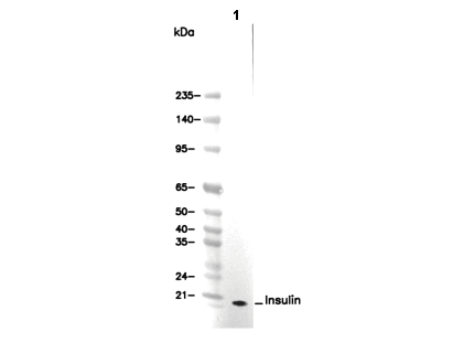 Lane 1: INS-1