
nur für Forschungszwecke
CUDC-101 HDAC Inhibitor
Kat.-Nr.: S1194
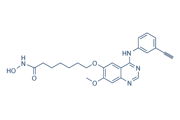
Chemische Struktur
Molekulargewicht: 434.49
Qualitätskontrolle (Quality Control)
Zellkultur, Behandlung & Arbeitskonzentration
(Cell Culture, Treatment & Working Concentration)
| Zelllinien | Assay-Typ | Konzentration | Inkubationszeit | Formulierung | Aktivitätsbeschreibung | PMID |
|---|---|---|---|---|---|---|
| human SK-BR-3 cells | Proliferation assay | Antiproliferative activity against human SK-BR-3 cells after hrs by ATP content assay, IC50=0.04 μM | ||||
| MDA-MB-231 cells | Proliferation assay | Antiproliferative activity against human MDA-MB-231 cells after hrs by ATP content assay, IC50=0.1 μM | ||||
| human HepG2 cells | Proliferation assay | Antiproliferative activity against human HepG2 cells after hrs by ATP content assay, IC50=0.13 μM | ||||
| human SKHEP1 cells | Proliferation assay | Antiproliferative activity against human SKHEP1 cells after hrs by ATP content assay, IC50=0.22 μM | ||||
| human Hep3B2 cells | Proliferation assay | Antiproliferative activity against human Hep3B2 cells after hrs by ATP content assay, IC50=0.23 μM | ||||
| human BxPC3 cells | Proliferation assay | Antiproliferative activity against human BxPC3 cells after hrs by ATP content assay, IC50=0.27 μM | ||||
| human NCI-H358 cells | Proliferation assay | Antiproliferative activity against human NCI-H358 cells after hrs by ATP content assay, IC50=0.4 μM | ||||
| human MCF7 cells | Proliferation assay | Antiproliferative activity against human MCF7 cells after hrs by ATP content assay, IC50=0.55 μM | ||||
| human HCC827 cells | Proliferation assay | Antiproliferative activity against human HCC827 cells after hrs by ATP content assay, IC50=0.6 μM | ||||
| human H460 cells | Proliferation assay | Antiproliferative activity against human H460 cells after hrs by ATP content assay, IC50=0.7 μM | ||||
| human Capan1 cells | Proliferation assay | Antiproliferative activity against human Capan1 cells after hrs by ATP content assay, IC50=0.8 μM | ||||
| Klicken Sie hier, um weitere experimentelle Daten zu Zelllinien anzuzeigen | ||||||
Chemische Informationen, Lagerung & Stabilität (Chemical Information, Storage & Stability)
| Molekulargewicht | 434.49 | Formel | C24H26N4O4 |
Lagerung (Ab dem Eingangsdatum) | |
|---|---|---|---|---|---|
| CAS-Nr. | 1012054-59-9 | SDF herunterladen | Lagerung von Stammlösungen |
|
|
| Synonyme | N/A | Smiles | COC1=C(C=C2C(=C1)N=CN=C2NC3=CC=CC(=C3)C#C)OCCCCCCC(=O)NO | ||
Löslichkeit (Solubility)
|
In vitro |
DMSO
: 43 mg/mL
(98.96 mM)
Water : Insoluble Ethanol : Insoluble |
Molaritätsrechner
|
In vivo |
|||||
In-vivo-Formulierungsrechner (Klare Lösung)
Schritt 1: Geben Sie die untenstehenden Informationen ein (Empfohlen: Ein zusätzliches Tier zur Berücksichtigung von Verlusten während des Experiments)
Schritt 2: Geben Sie die In-vivo-Formulierung ein (Dies ist nur der Rechner, keine Formulierung. Bitte kontaktieren Sie uns zuerst, wenn es im Abschnitt "Löslichkeit" keine In-vivo-Formulierung gibt.)
Berechnungsergebnisse:
Arbeitskonzentration: mg/ml;
Methode zur Herstellung der DMSO-Stammlösung: mg Wirkstoff vorgelöst in μL DMSO ( Konzentration der Stammlösung mg/mL, Bitte kontaktieren Sie uns zuerst, wenn die Konzentration die DMSO-Löslichkeit der Wirkstoffcharge überschreitet. )
Methode zur Herstellung der In-vivo-Formulierung: Nehmen Sie μL DMSO Stammlösung, dann hinzufügenμL PEG300, mischen und klären, dann hinzufügenμL Tween 80, mischen und klären, dann hinzufügen μL ddH2O, mischen und klären.
Methode zur Herstellung der In-vivo-Formulierung: Nehmen Sie μL DMSO Stammlösung, dann hinzufügen μL Maisöl, mischen und klären.
Hinweis: 1. Bitte stellen Sie sicher, dass die Flüssigkeit klar ist, bevor Sie das nächste Lösungsmittel hinzufügen.
2. Achten Sie darauf, das/die Lösungsmittel der Reihe nach hinzuzufügen. Sie müssen sicherstellen, dass die bei der vorherigen Zugabe erhaltene Lösung eine klare Lösung ist, bevor Sie mit der Zugabe des nächsten Lösungsmittels fortfahren. Physikalische Methoden wie Vortex, Ultraschall oder ein heißes Wasserbad können zur Unterstützung des Lösens verwendet werden.
Wirkmechanismus (Mechanism of Action)
| Targets/IC50/Ki |
EGFR
(Cell-free assay) 2.4 nM
HDAC
(Cell-free assay) 4.4 nM
HDAC1
(Cell-free assay) 4.5 nM
HDAC6
(Cell-free assay) 5.1 nM
HDAC3
(Cell-free assay) 9.1 nM
HDAC5
(Cell-free assay) 11.4 nM
HDAC2
(Cell-free assay) 12.6 nM
HDAC4
(Cell-free assay) 13.2 nM
HER2
(Cell-free assay) 15.7 nM
HDAC10
(Cell-free assay) 26.1 nM
HDAC9
(Cell-free assay) 67.2 nM
HDAC8
(Cell-free assay) 79.8 nM
HDAC7
(Cell-free assay) 373 nM
|
|---|---|
| In vitro |
Specific for class I and class II HDACs, CUDC-101 does not inhibit class III Sir-type HDACs. This compound displays weak activity against other protein kinases including KDR/VEGFR2, Lyn, Lck, Abl-1, FGFR-2, Flt-3, and Ret with IC50 of 0.85 μM, 0.84 μM, 5.91 μM, 2.89 μM, 3.43 μM, 1.5 μM, abd 3.2 μM, respectively. It displays broad antiproliferative activity in many human cancer cell types with IC50 of 0.04-0.80 μM, exhibiting a higher potency and combinations of vorinostat in most cases. This chemical potently inhibits cancer cell lines. It inhibits the resistant EGFR mutant T790M although its effects are incomplete with an Amax of ~60% of peak enzyme activity after inhibition. This compound treatment increases the acetylation of histone H3 and H4, as well as the acetylation of non-histone substrates of HDAC such as p53 and α-tubulin, in a dose-dependant manner in various cancer cell lines. It also suppresses HER3 expression, Met amplification, and AKT reactivation in tumor cells. |
| Kinase-Assay |
HDAC-, EGFR- und HER2-Hemmungstests
|
|
Die Aktivitäten von HDACs der Klasse I und II werden mit dem Biomol Color de Lys System beurteilt. Kurz gesagt, werden HeLa-Zellkernextrakte als Quelle für HDACs verwendet. Verschiedene Konzentrationen von CUDC-101 werden zu HeLa-Zellkernextrakten in Gegenwart eines kolorimetrischen künstlichen Substrats hinzugefügt. Am Ende des Assays wird Entwickler hinzugefügt und die Enzymaktivität im Wallac Victor II 1420 Mikroplattenleser bei 405 nM gemessen. Die EGFR- und HER2-Kinaseaktivität wird mit HTScan EGF-Rezeptor- und HER2-Kinase-Assay-Kits gemessen. Kurz gesagt, das GST-EGFR-Fusionsprotein wird mit einem synthetischen biotinylierten Peptidsubstrat und variierenden Konzentrationen dieser Verbindung in Gegenwart von 400 mM ATP inkubiert. Phosphoryliertes Substrat wird mit Streptavidin-beschichteten 96-Well-Platten eingefangen. Der Grad der Phosphorylierung wird durch anti-Phosphotyrosin- und Europium-markierte Sekundärantikörper überwacht. Am Ende des Assays wird die Verstärkungslösung hinzugefügt und die Enzymaktivität im Wallac Victor II 1420 Mikroplattenleser bei 615 nM gemessen.
|
|
| In vivo |
Administration of CUDC-101 at 120 mg/kg/day induces tumor regression in the Hep-G2 liver cancer model, which is more efficacious at its maximum tolerated dose (25 mg/kg/day) and vorinostat at an equimolar concentration dose (72 mg/kg/day). This compound inhibits the growth of -sensitive H358 NSCLC xenografts in a dose-dependent manner. It also shows potent inhibition of tumor growth in the -resistant A549 NSCLC xenograft model. This chemical produces significant tumor regression in the -resistant, HER2-negative, EGFR-overexpressing MDA-MB-468 breast cancer model and the EGFR-overexpressing CAL-27 head and neck squamous cell carcinoma (HNSCC) model. Additionally, it inhibits tumor growth in the K-ras mutant HCT116 colorectal and EGFR/HER2 (neu)-expressing HPAC pancreatic cancer models. |
Literatur |
|
Klinische Studieninformationen (Clinical Trial Information)
(Daten von https://clinicaltrials.gov, aktualisiert am 2018-02-22)
| NCT-Nummer | Rekrutierung | Erkrankungen | Sponsor/Kooperationspartner | Startdatum | Phasen |
|---|---|---|---|---|---|
| NCT01384799 | COMPLETED | Head and Neck Cancer |
Curis, Inc. |
2011-11 | PHASE1 |
| NCT01702285 | TERMINATED | Cancer |
Curis, Inc. |
2012-09 | PHASE1 |
| NCT01702285 | Terminated | Cancer |
Curis Inc. |
2012-09 | Phase 1 |
| NCT01384799 | Completed | Head and Neck Cancer |
Curis Inc. |
2011-11 | Phase 1 |
| NCT01171924 | COMPLETED | Head and Neck Cancer; Liver Cancer; Breast Cancer; Gastric Cancer; Non-Small Cell Lung Cancer |
Curis, Inc. |
2010-07 | PHASE1 |
| NCT00728793 | COMPLETED | Tumors |
Curis, Inc. |
2008-08 | PHASE1 |

Produkte sind nur für Forschungszwecke bestimmt. Nicht für den menschlichen Gebrauch. Wir verkaufen nicht an Patienten.
©Copyright 2013 Selleck Chemicals. Alle Rechte vorbehalten.