|
Wie zu zitieren 1. Für Zitate im Text (Materialien & Methoden): 2. Für die Tabelle der Schlüsselressourcen: |
||
|
Gebührenfrei: (877) 796-6397 -- Nur USA und Kanada -- |
Fax: +1-832-582-8590 Bestellungen: +1-832-582-8158 |
Technischer Support: +1-832-582-8158 Ext:3 Bitte geben Sie Ihre Bestellnummer in der E-Mail an. Wir bemühen uns, alle E-Mail-Anfragen innerhalb eines Werktages zu beantworten. |
Biologische Beschreibung
| Spezifität | 8-Hydroxy-2'-deoxyguanosine Antibody [K11E8] weist sowohl endogenes als auch exogenes 8-Hydroxy-2'-deoxyguanosin (8-OH-dG) nach. |
|---|---|
| Hintergrund | 8-Hydroxy-2'-deoxyguanosin (8-OHdG) ist ein modifiziertes Purin-2'-desoxyribonukleosid, das durch die Hydroxylierung der Guaninbase an der C8-Position in der DNA entsteht, typischerweise infolge oxidativer Schäden, die durch reaktive Sauerstoffspezies (ROS) verursacht werden. Strukturell besteht es aus einem Desoxyguanosin-Molekül mit einer Hydroxylgruppe am 8. Kohlenstoff des Guaninrings. 8-OHdG ist aufgrund seiner hohen Prävalenz unter oxidativen DNA-Läsionen weithin als wichtiger Biomarker für oxidativen Stress anerkannt. Es reichert sich sowohl in der nuklearen als auch in der mitochondrialen DNA an und kann in verschiedenen biologischen Proben gemessen werden, einschließlich Urin, Geweben und Leukozyten-DNA. Funktionell dient 8-OHdG als Indikator für endogene DNA-Schäden und wurde ausgiebig verwendet, um das Risiko und die Progression von Krebs, Alterung und degenerativen Erkrankungen zu bewerten. Seine Anwesenheit spiegelt die zelluläre Exposition gegenüber oxidativen Agenzien wider und ist maßgeblich an der Bewertung der Auswirkungen von Umwelt- und Lebensstilfaktoren auf die genomische Stabilität beteiligt. |
Nutzungsinformationen
| Anwendung | IHC-P | Verdünnung |
|
||
|---|---|---|---|---|---|
| Reaktivität | Species independent | ||||
| Quelle | Mouse Monoclonal Antibody | MW | |||
| Lagerpuffer | PBS, pH 7.2+50% Glycerol+0.05% BSA+0.01% NaN3 | Lagerung (Ab dem Datum des Erhalts) |
-20°C (avoid freeze-thaw cycles), 2 years | ||
| IHC |
Experimental Protocol:
Deparaffinization/Rehydration
1. Deparaffinize/hydrate sections:
2. Incubate sections in three washes of xylene for 5 min each.
3. Incubate sections in two washes of 100% ethanol for 10 min each.
4. Incubate sections in two washes of 95% ethanol for 10 min each.
5. Wash sections two times in dH2O for 5 min each.
6.Antigen retrieval: For Citrate: Heat slides in a microwave submersed in 1X citrate unmasking solution until boiling is initiated; continue with 10 min at a sub-boiling temperature (95°-98°C). Cool slides on bench top for 30 min.
Staining
1. Wash sections in dH2O three times for 5 min each.
2. Incubate sections in 3% hydrogen peroxide for 10 min.
3. Wash sections in dH2O two times for 5 min each.
4. Wash sections in wash buffer for 5 min.
5. Block each section with 100–400 µl of blocking solution for 1 hr at room temperature.
6. Remove blocking solution and add 100–400 µl primary antibody diluent in to each section. Incubate overnight at 4°C.
7. Remove antibody solution and wash sections with wash buffer three times for 5 min each.
8. Cover section with 1–3 drops HRPas needed. Incubate in a humidified chamber for 30 min at room temperature.
9. Wash sections three times with wash buffer for 5 min each.
10. Add DAB Chromogen Concentrate to DAB Diluent and mix well before use.
11. Apply 100–400 µl DAB to each section and monitor closely. 1–10 min generally provides an acceptable staining intensity.
12. Immerse slides in dH2O.
13. If desired, counterstain sections with hematoxylin.
14. Wash sections in dH2O two times for 5 min each.
15. Dehydrate sections: Incubate sections in 95% ethanol two times for 10 sec each; Repeat in 100% ethanol, incubating sections two times for 10 sec each; Repeat in xylene, incubating sections two times for 10 sec each.
16. Mount sections with coverslips and mounting medium.
|
Referenzen
|
Anwendungsdaten
IHC
Validiert von Selleck
-
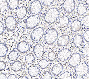 Immunohistochemical analysis of formalin fixed paraffin embedded human colorectal cancer tissue with F1714 at 1:50 dilution.