Technische Daten
| Formel | C29H26ClFN4O4S.2C7H8O3S |
||||||
| Molekulargewicht | 925.46 | CAS-Nr. | 388082-77-7 | ||||
| Löslichkeit (25°C)* | In vitro | DMSO | 100 mg/mL (108.05 mM) | ||||
| Water | Insoluble | ||||||
| Ethanol | Insoluble | ||||||
| In vivo (Lösungsmittel einzeln und der Reihe nach zum Produkt hinzufügen.) |
|
||||||
|
* <1 mg/ml bedeutet schwer löslich oder unlöslich. * Bitte beachten Sie, dass Selleck die Löslichkeit aller Verbindungen intern testet und die tatsächliche Löslichkeit geringfügig von veröffentlichten Werten abweichen kann. Dies ist normal und ist auf geringfügige Batch-zu-Batch-Variationen zurückzuführen. * Versand bei Raumtemperatur (Stabilitätstests zeigen, dass dieses Produkt ohne Kühlmaßnahmen versendet werden kann.) |
|||||||
Vorbereitung von Stammlösungen
Biologische Aktivität
| Beschreibung | Lapatinib Ditosylate ist ein potenter EGFR- und ErbB2-Inhibitor mit einer IC50 von 10,8 bzw. 9,2 nM in zellfreien Assays. | ||||||
|---|---|---|---|---|---|---|---|
| Ziele |
|
||||||
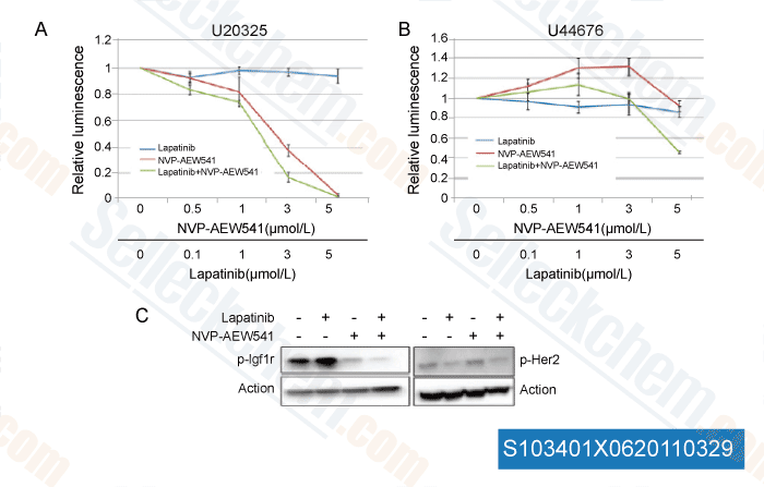
| In vitro | Lapatinib Ditosylate hemmt schwach die Aktivität von ErbB4 mit einer IC50 von 367 nM und zeigt eine >300-fache Selektivität für EGFR und ErbB2 gegenüber anderen Kinasen wie c-Src, c-Raf, MEK, ERK, c-Fms, CDK1, CDK2, p38, Tie-2 und VEGFR2. Diese Verbindung hemmt signifikant die Rezeptorautophosphorylierung von EGFR und ErbB2 dosisabhängig mit einer IC50 von 170 nM bzw. 80 nM in HN5-Zellen; sowie 210 nM bzw. 60 nM in BT474-Zellen. Im Gegensatz zu OSI-774 und Iressa (ZD1839), die bevorzugt das Wachstum von EGFR-überexprimierenden Zellen hemmen, hemmt es das Wachstum sowohl von EGFR- als auch von ErbB2-überexprimierenden Zellen. Es zeigt eine höhere Hemmaktivität gegen EGFR- oder ErbB2-überexprimierende Zellen mit einer IC50 von 0,09-0,21 μM, verglichen mit Zellen, die niedrige Spiegel von EGFR oder ErbB2 exprimieren, mit einer IC50 von 3-12 μM, und weist eine ~100-fache Selektivität gegenüber normalen Fibroblastenzellen auf. Diese Chemikalie hemmt potent das Auswachsen von EGFR-überexprimierenden HN5- und A-431-Zellen sowie von ErbB2-überexprimierenden BT474- und N87-Zellen und induziert signifikant G1-Arrest in HN5-Zellen und Apoptose in BT474-Zellen, was mit der Hemmung der AKT-Phosphorylierung verbunden ist. | ||||||
| In vivo | Die orale Verabreichung von Lapatinib Ditosylate (~100 mg/kg) zweimal täglich hemmt signifikant das Wachstum von BT474- und HN5-Xenotransplantaten dosisabhängig. |
Protokoll (aus Referenz)
| Kinase-Assay:[1] |
|
|---|---|
| Zell-Assay:[1] |
|
| Tierstudie:[1] |
|
Referenzen
|
Kundenproduktvalidierung
-
Daten von [ Mol Cancer Ther , 2011 , 10:697-707 ]
-
Daten von [ Biochem Pharmacol , 2011 , 82, 1457-1466 ]
-
Daten von [ Biochem Pharmacol , 2011 , 82, 1457-1466 ]
-
Daten von [ Int J Proteomics , 2011 , 2011, 215496 ]
Sellecks Lapatinib Ditosylate Wurde zitiert von 149 Publikationen
| Inactivation of necroptosis-promoting protein MLKL creates a therapeutic vulnerability in colorectal cancer cells [ Cell Death Dis, 2025, 16(1):118] | PubMed: 39979285 |
| Multiscale Modeling Uncovers Macrophage Infiltration and TNF-α Signaling Networks for Targeting in Inflammatory Breast Cancer Tumor Emboli [ bioRxiv, 2025, 2025.05.29.656249] | PubMed: 40502021 |
| Altered ribosomal profile in acquired resistance and reversal associates with pathological response to chemotherapy in inflammatory breast cancer [ NPJ Breast Cancer, 2024, 10(1):65] | PubMed: 39075068 |
| Patient-derived rhabdomyosarcoma cells recapitulate the genetic and transcriptomic landscapes of primary tumors [ iScience, 2024, 27(10):110862] | PubMed: 39319271 |
| Profiling of ERBB receptors and downstream pathways reveals selectivity and hidden properties of ERBB4 antagonists [ iScience, 2024, 27(2):108839] | PubMed: 38303712 |
| Massively parallel reporter assays identify enhancer elements in oesophageal Adenocarcinoma [ NAR Cancer, 2024, 6(4):zcae041] | PubMed: 39417090 |
| Overcoming brain-derived therapeutic resistance in HER2+ breast cancer brain metastasis [ bioRxiv, 2024, 2024.02.19.581073] | PubMed: 38529509 |
| Proteomic Assessment of SKBR3/HER2+ Breast Cancer Cellular Response to Lapatinib and Investigational Ipatasertib Kinase Inhibitors [ bioRxiv, 2024, 2024.04.02.587656] | PubMed: 38617302 |
| Protocol for identifying properties of ERBB receptor antagonists using the barcoded ERBBprofiler assay [ STAR Protoc, 2024, 5(2):102987] | PubMed: 38635397 |
| Analysis and modeling of cancer drug responses using cell cycle phase-specific rate effects [ Nat Commun, 2023, 14(1):3450] | PubMed: 37301933 |
RÜCKGABERICHTLINIE
Die bedingungslose Rückgaberichtlinie von Selleck Chemical gewährleistet unseren Kunden ein reibungsloses Online-Einkaufserlebnis. Wenn Sie in irgendeiner Weise mit Ihrem Kauf unzufrieden sind, können Sie jeden Artikel innerhalb von 7 Tagen nach Erhalt zurückgeben. Im Falle von Produktqualitätsproblemen, sei es protokollbezogene oder produktbezogene Probleme, können Sie jeden Artikel innerhalb von 365 Tagen ab dem ursprünglichen Kaufdatum zurückgeben. Bitte befolgen Sie die nachstehenden Anweisungen, wenn Sie Produkte zurücksenden.
VERSAND UND LAGERUNG
Selleck-Produkte werden bei Raumtemperatur transportiert. Wenn Sie das Produkt bei Raumtemperatur erhalten, seien Sie versichert, dass die Qualitätskontrollabteilung von Selleck Experimente durchgeführt hat, um zu überprüfen, dass die normale Temperaturplatzierung von einem Monat die biologische Aktivität von Pulverprodukten nicht beeinträchtigt. Nach dem Sammeln lagern Sie das Produkt bitte gemäß den in der Datenblatt beschriebenen Anforderungen. Die meisten Selleck-Produkte sind unter den empfohlenen Bedingungen stabil.
NICHT FÜR DIE ANWENDUNG AM MENSCHEN, FÜR VETERINÄRMEDIZINISCHE DIAGNOSTIK ODER THERAPEUTISCHE ZWECKE.