Technische Daten
| Formel | C17H15FN2O3 |
||||||
| Molekulargewicht | 314.31 | CAS-Nr. | 852475-26-4 | ||||
| Löslichkeit (25°C)* | In vitro | DMSO | 13 mg/mL (41.36 mM) | ||||
| Water | Insoluble | ||||||
| Ethanol | Insoluble | ||||||
| In vivo (Lösungsmittel einzeln und der Reihe nach zum Produkt hinzufügen.) |
|
||||||
|
* <1 mg/ml bedeutet schwer löslich oder unlöslich. * Bitte beachten Sie, dass Selleck die Löslichkeit aller Verbindungen intern testet und die tatsächliche Löslichkeit geringfügig von veröffentlichten Werten abweichen kann. Dies ist normal und ist auf geringfügige Batch-zu-Batch-Variationen zurückzuführen. * Versand bei Raumtemperatur (Stabilitätstests zeigen, dass dieses Produkt ohne Kühlmaßnahmen versendet werden kann.) |
|||||||
Vorbereitung von Stammlösungen
Biologische Aktivität
| Beschreibung | MC1568 ist ein selektiver HDAC-Inhibitor für Mais-HD1-A mit einem IC50-Wert von 100 nM in einem zellfreien Assay. Er ist 34-fach selektiver für HD1-A als für HD1-B. | ||||
|---|---|---|---|---|---|
| Ziele |
|
||||
| In vitro | MC1568 ist ein selektiver Histon-Deacetylase (HDAC II)-Inhibitor der Klasse II (IIa) mit einem IC50 von 220 nM und einer 176-fachen Selektivität der Klasse II (gegenüber Klasse I). In Lysaten menschlicher Brustkrebs-ZR-75.1-Zellen zeigt diese Verbindung (5 μM) keine hemmende Wirkung gegen HDAC1, kann aber HDAC4 hemmen. In MCF-7-Zellen erhöht diese Verbindung (20 μM) die Akkumulation von acetylierten H3- und H4-Histonen sowie die Spiegel von Acetyl-Tubulin, was auf eine hemmende Wirkung dieser Chemikalie auf HDAC6 hinweist. In C2C12-Zellen hemmt diese Verbindung (5 μM) die Myogenese, indem sie die Expression des Myozyten-Enhancer-Faktors 2D (MEF2D) verringert, den HDAC4-HDAC3-MEF2D-Komplex stabilisiert und die differenzierungsinduzierte MEF2D-Acetylierung hemmt. Diese Verbindung (5 oder 10 μM) interferiert mit den RAR- und PPARγ-vermittelten differenzierungsinduzierenden Signalwegen. In F9-Zellen blockiert diese Chemikalie spezifisch die endodermale Differenzierung, obwohl sie die Retinsäure-induzierte Reifung von promyelozytischen NB4-Zellen nicht beeinflusst. In 3T3-L1-Zellen schwächt diese Verbindung die PPARγ-induzierte Adipogenese ab. |
||||
| In vivo | Bei Mäusen zeigt MC1568 (50 mg/kg) eine offensichtliche gewebeselektive HDAC-Hemmung. Im Skelettmuskel und Herzen hemmt diese Verbindung die Aktivität von HDAC4 und HDAC5, ohne die HDAC3-Aktivität zu beeinflussen, wodurch MEF2-HDAC-Komplexe in einem reprimierten Zustand verbleiben. Bei PPRE-Luc-Mäusen beeinträchtigt es die PPARγ-Signalübertragung hauptsächlich im Herzen und im Fettgewebe. In einer kürzlich durchgeführten Studie an Pankreasexplantaten verstärkt diese Chemikalie die Expression von Pax4, einem Schlüsselfaktor, der für die ordnungsgemäße β- und δ-Zelldifferenzierung erforderlich ist, und amplifiziert endokrine β- und δ-Zellen. |
Protokoll (aus Referenz)
| Kinase-Assay: |
|
|---|---|
| Zell-Assay: |
|
| Tierstudie: |
|
Referenzen
|
Kundenproduktvalidierung

-
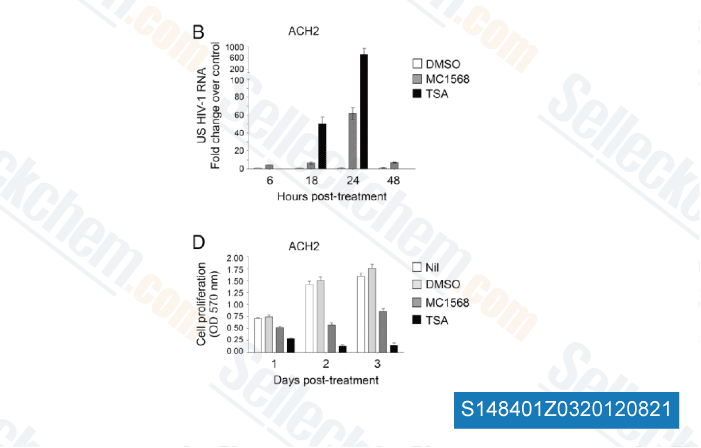
Daten von [ Proc Natl Acad Sci U S A , 2012 , 109(34) ]
-
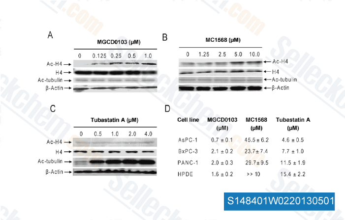
Daten von [ Proc Natl Acad Sci U S A , 2012 , 109(34) ]
-
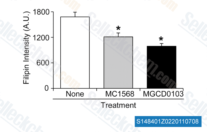
Daten von [ PLoS One , 2012 , 7(12), e52095 ]
-
Daten von [ J Biol Chem , 2011 , 286, 23842–23851 ]
Sellecks MC1568 Wurde zitiert von 51 Publikationen
| HSD17B4 deficiency causes dysregulation of primary cilia and is alleviated by acetyl-CoA [ Nat Commun, 2025, 16(1):2663] | PubMed: 40102401 |
| Key epigenetic and signaling factors in the formation and maintenance of the blood-brain barrier [ Elife, 2024, 12RP86978] | PubMed: 39670988 |
| Global isonicotinylome analysis identified SMAD3 isonicotinylation promotes liver cancer cell epithelial-mesenchymal transition and invasion [ iScience, 2024, 27(9):110775] | PubMed: 39286495 |
| Selective Inhibition of Histone Deacetylase Class IIa With MC1568 Ameliorates Podocyte Injury [ Front Med (Lausanne), 2022, 9:848938] | PubMed: 35492337 |
| HBV covalently closed circular DNA minichromosomes in distinct epigenetic transcriptional states differ in their vulnerability to damage [ Hepatology, 2021, 10.1002/hep.32245] | PubMed: 34779008 |
| Butyrate and Class I Histone Deacetylase Inhibitors Promote Differentiation of Neonatal Porcine Islet Cells into Beta Cells [ Cells, 2021, 10(11)3249] | PubMed: 34831471 |
| Epigenetic Repression of Chloride Channel Accessory 2 Transcription in Cardiac Fibroblast: Implication in Cardiac Fibrosis [ Front Cell Dev Biol, 2021, 9:771466] | PubMed: 34869368 |
| HDAC8 cooperates with SMAD3/4 complex to suppress SIRT7 and promote cell survival and migration. [ Nucleic Acids Res, 2020, 6;48(6):2912-2923] | PubMed: 31970414 |
| Valproate reverses mania-like behaviors in mice via preferential targeting of HDAC2 [ Mol Psychiatry, 2020, 10.1038/s41380-020-00958-2] | PubMed: 33235333 |
| Valproic acid upregulates the expression of the p75NTR/sortilin receptor complex to induce neuronal apoptosis [ Apoptosis, 2020, 10.1007/s10495-020-01626-0] | PubMed: 32712736 |
RÜCKGABERICHTLINIE
Die bedingungslose Rückgaberichtlinie von Selleck Chemical gewährleistet unseren Kunden ein reibungsloses Online-Einkaufserlebnis. Wenn Sie in irgendeiner Weise mit Ihrem Kauf unzufrieden sind, können Sie jeden Artikel innerhalb von 7 Tagen nach Erhalt zurückgeben. Im Falle von Produktqualitätsproblemen, sei es protokollbezogene oder produktbezogene Probleme, können Sie jeden Artikel innerhalb von 365 Tagen ab dem ursprünglichen Kaufdatum zurückgeben. Bitte befolgen Sie die nachstehenden Anweisungen, wenn Sie Produkte zurücksenden.
VERSAND UND LAGERUNG
Selleck-Produkte werden bei Raumtemperatur transportiert. Wenn Sie das Produkt bei Raumtemperatur erhalten, seien Sie versichert, dass die Qualitätskontrollabteilung von Selleck Experimente durchgeführt hat, um zu überprüfen, dass die normale Temperaturplatzierung von einem Monat die biologische Aktivität von Pulverprodukten nicht beeinträchtigt. Nach dem Sammeln lagern Sie das Produkt bitte gemäß den in der Datenblatt beschriebenen Anforderungen. Die meisten Selleck-Produkte sind unter den empfohlenen Bedingungen stabil.
NICHT FÜR DIE ANWENDUNG AM MENSCHEN, FÜR VETERINÄRMEDIZINISCHE DIAGNOSTIK ODER THERAPEUTISCHE ZWECKE.