OSI-930

Katalog-Nr.S1220 Charge:S122001

Drucken

Technische Daten

Formel

C22H16F3N3O2S

Molekulargewicht 443.44 CAS-Nr. 728033-96-3
Löslichkeit (25°C)* In vitro DMSO 89 mg/mL (200.7 mM)
Ethanol 3 mg/mL (6.76 mM)
Water Insoluble
In vivo (Lösungsmittel einzeln und der Reihe nach zum Produkt hinzufügen.)
Homogeneous suspension
CMC-NA
≥5mg/ml Taking the 1 mL working solution as an example, add 5 mg of this product to 1 ml of CMC-Na solution, mix evenly to obtain a homogeneous suspension with a final concentration of 5 mg/ml.
* <1 mg/ml bedeutet schwer löslich oder unlöslich.
* Bitte beachten Sie, dass Selleck die Löslichkeit aller Verbindungen intern testet und die tatsächliche Löslichkeit geringfügig von veröffentlichten Werten abweichen kann. Dies ist normal und ist auf geringfügige Batch-zu-Batch-Variationen zurückzuführen.
* Versand bei Raumtemperatur (Stabilitätstests zeigen, dass dieses Produkt ohne Kühlmaßnahmen versendet werden kann.)

Vorbereitung von Stammlösungen

Biologische Aktivität

Beschreibung OSI-930 ist ein potenter Inhibitor von Kit (c-Kit), KDR und CSF-1R mit IC50 von 80 nM, 9 nM bzw. 15 nM; diese Verbindung ist auch potent gegenüber Flt-1, c-Raf und Lck und hat eine geringe Aktivität gegen PDGFRα/β, Flt-3 und Abl. Phase 1.
Ziele
FLT1
(Cell-free assay)
KDR
(Cell-free assay)
CSF-1R
(Cell-free assay)
LCK
(Cell-free assay)
C-Raf
(Cell-free assay)
Mehr anzeigen
8 nM 9 nM 15 nM 22 nM 41 nM
In vitro OSI-930 hemmt die Zellproliferation in der HMC-1-Zelllinie mit einer IC50 von 14 nM ohne signifikanten Einfluss auf das Wachstum der COLO-205-Zelllinie, die keinen konstitutiv aktiven mutierten Rezeptor-Tyrosinkinase exprimiert. Darüber hinaus induziert diese Verbindung auch die Apoptose in der HMC-1-Zelllinie mit einer EC50 von 34 nM. Eine kürzlich durchgeführte Studie zeigt, dass diese Chemikalie gereinigtes, rekombinantes Cytochrom P450 (P450) 3A4 mit einem Ki von 24 μM zeit- und konzentrationsabhängig inaktiviert.
In vivo OSI-930, oral verabreicht in der maximal wirksamen Dosis von 200 mg/kg mittels Magensonde, zeigt eine potente Antitumoraktivität in einem breiten Spektrum präklinischer Xenograft-Modelle, einschließlich HMC-1, NCI-SNU-5, COLO-205 und U251 Xenograft-Modelle.

Protokoll (aus Referenz)

Kinase-Assay:[1]
  • Proteinkinase-Assays

    Proteinkinase-Assays werden entweder intern mittels ELISA-basierter Assay-Methoden (Kit, KDR, PDGFRα und PDGFRβ) oder mittels einer radiometrischen Methode durchgeführt. Die internen ELISA-Assays verwendeten Poly(Glu:Tyr) als Substrat, gebunden an die Oberfläche von 96-Well-Assay-Platten; die Phosphorylierung wird dann unter Verwendung eines Antiphosphotyrosin-Antikörpers, konjugiert an HRP, nachgewiesen. Der gebundene Antikörper wird dann unter Verwendung von ABTS als Peroxidase-Substrat durch Messung der Absorption bei 405/490 nm quantifiziert. Alle Assays verwenden gereinigte rekombinante Kinase-katalytische Domänen, die entweder in Insektenzellen oder in Bakterien exprimiert werden. Das für interne Assays verwendete Kit- und EGFR-Protein wird intern hergestellt; andere Enzyme werden bezogen. Rekombinantes Kit-Protein wird als NH2-terminales Glutathion-S-Transferase-Fusionsprotein in Insektenzellen exprimiert und ist initial als nicht-phosphoryliertes (nicht-aktiviertes) Enzym mit einem relativ hohen Km für ATP (400 μM) gereinigt. In einigen Assays wird eine aktivierte (Tyrosin-phosphorylierte) Form des Enzyms durch Inkubation mit 1 mM ATP für 1 Stunde bei 30 °C hergestellt. Das phosphorylierte Protein wird dann durch eine Entsalzungssäule geleitet, um den Großteil des ATP zu entfernen und bei –80 °C in Puffer mit 50 % Glycerin gelagert. Das resultierende Präparat hat eine deutlich höhere spezifische Aktivität und einen niedrigeren Km für ATP (25 μM) als das initiale nicht-phosphorylierte Präparat. Die Hemmung der Kit-Autophosphorylierung durch diese Verbindung wird durch Inkubation des nicht-phosphorylierten Enzyms bei 30 °C in Gegenwart von 200 μM ATP und verschiedenen Konzentrationen dieser Chemikalie untersucht. Die Reaktion wird durch Entnahme von Aliquoten in SDS-PAGE-Probenpuffer und anschließendes Erhitzen auf 100 °C für 5 Minuten gestoppt. Der Grad der Phosphorylierung von Kit wird dann durch Immunoblotting für sowohl Gesamt-Kit als auch phosphoryliertes Kit bestimmt.

Zell-Assay:[1]
  • Zelllinien

    HMC-1 and COLO-205

  • Konzentrationen

    0--1 μM

  • Inkubationszeit

    48 hours

  • Methode

    For assays of cell proliferation and apoptosis, cells are seeded into 96-well plates and incubated for 2 to 3 days in the presence of OSI-930 at various concentrations. Inhibition of cell growth is determined by luminescent quantitation of the intracellular ATP content using CellTiterGlo. Induction of caspase-dependent apoptosis by this compound is quantitated by an enzymatic caspase 3/7 assay. Inhibition of angiogenesis by this chemical is monitored using the rat aortic ring endothelial sprout outgrowth assay. Sections of aorta are prepared from CO2-euthanized male rats and cultured in vitro in a collagen matrix in the presence or absence of this compound. The collagen matrix is prepared from type 1 rat tail collagen solubilized in 0.1% acetic acid at 3 mg/mL, which is combined with 0.125 volume collagen buffer (0.05 N NaOH, 200 mM HEPES, 260 mM NaHCO3), 0.125 volume of medium 199, 0.0125 volume of 1 M NaOH, and 1% GlutaMax. Aortic rings are embedded in 0.4 mL of this matrix in six-well plates, to which 0.5 mL endothelial basal medium and the appropriate amount of this chemical is added; the rings are then incubated for 10 days and the resultant angiogenic sprout outgrowth is digitally quantitated from images by measurement of the sprout-containing area within a series of concentric rings around the aortic tissue area.

Tierstudie:[1]
  • Tiermodelle

    HMC-1, NCI-SNU-5, COLO-205 and U251 cells are injected s.c. into the right flank of CD1 nu/nu mice.

  • Dosierungen

    ≤200 mg/kg

  • Verabreichung

    Administered via p.o.

Referenzen

  • https://pubmed.ncbi.nlm.nih.gov/16424037/
  • https://pubmed.ncbi.nlm.nih.gov/21068193/

Kundenproduktvalidierung

RE-luc2P-HEK293 cells were pretreated with 1uM OSI-930 (green), 20uM TBB (blue), 10uM CKI-7 (purple), or 10uM H-89 (orange) for 16 h and infected with Y. enterocolitica WA or Y. pestis Ind195 at MOI 1 and 20, respectively, for 1 h. Following stimulation with 10 ng/ml TNF-α at 5 h post-infection, luciferase activity was measured 24 h post-infection. Results were determined from two independent experiments performed in triplicate. A"*" denotes that the % NF-κβ inhibition using the inhibitors was significantly different (p<0.05) compared to the no drug control (black). The relative NF-κB inhibition by Yersinia infection was determined as a percentage of luciferase activity in bacteria-infected cells relative to luciferase activity in bacteria-free control cells.

Daten von [ BMC Microbiol , 2013 , 13, 249 ]

<p> </p><p>Table 2. shows the reversal effect of OSI-930 and Fumitremorgin C (FTC) on the cytotoxicity of mitoxantrone to HEK293/pcDNA3.1 and HEK293-ABCG2-482-R2.<sup>a</sup>IC<sub>50</sub>: concentration that inhibited cell survival by 50% (means ± SD). <sup>b</sup>FR: fold-resistance was the value of that IC<sub>50</sub> value for mitoxantrone of HEK293/pcDNA3.1 cells was divided by IC<sub>50</sub> value for mitoxantrone of HEK293/pcDNA3.1 and HEK293-ABCG2-482-R2 cells in the absence or presence of OSI-930 and Fumitremorgin C (FTC). Values in table are representative of at least three independent experiments performed in triplicate.</p>

,

<p>For MTT assays, cells (2,000 ~ 5,000 cells/well) were subcultured into 96-well plates according to their growth properties. Cell proliferation was assayed at 72 hr after treatment of OSI-930 by adding 20 μl of 5 mg/ml 3-(4,5-Dimethylthiazol-2-yl)-2,5-diphenyltetrazolium bromide (MTT) solution per 100 μl of growth medium. After incubating for 3-4 h at 37°C, the media were removed and 150 µl/well of MTT solvent (either absolute DMSO or isopropanol containing 4 mM HCl and 0.1% Nonidet-40) was added to dissolve the formazan</p>

, , Dr. Yong-Weon Yi from Georgetown University Medical Center

Sellecks OSI-930 Wurde zitiert von 7 Publikationen

Orthogonal proteogenomic analysis identifies the druggable PA2G4-MYC axis in 3q26 AML [ Nat Commun, 2024, 15(1):4739] PubMed: 38834613
Small-Molecule and CRISPR Screening Converge to Reveal Receptor Tyrosine Kinase Dependencies in Pediatric Rhabdoid Tumors. [ Cell Rep, 2019, 28(9):2331-2344] PubMed: 31461650
TLR7/8-agonist-loaded Nanoparticles Promote the Polarization of Tumour-Associated Macrophages to Enhance Cancer Immunotherapy [ Nat Biomed Eng, 2018, 2(8):578-588] PubMed: 31015631
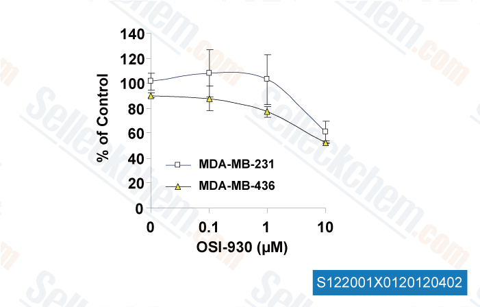
Targeting a cell state common to triple-negative breast cancers [Muellner MK, et al. Mol Syst Biol, 2015, 11(1):789] PubMed: 25699542
Dual inhibition of EGFR and MET induces synthetic lethality in triple-negative breast cancer cells through downregulation of ribosomal protein S6. [Yi YW, et al. Int J Oncol, 2015, 47(1):122-32] PubMed: 25955731
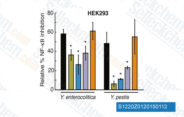
c-KIT signaling is targeted by pathogenic Yersinia to suppress the host immune response. [Micheva-Viteva SN, et al. BMC Microbiol, 2013, 13(1):249] PubMed: 24206648
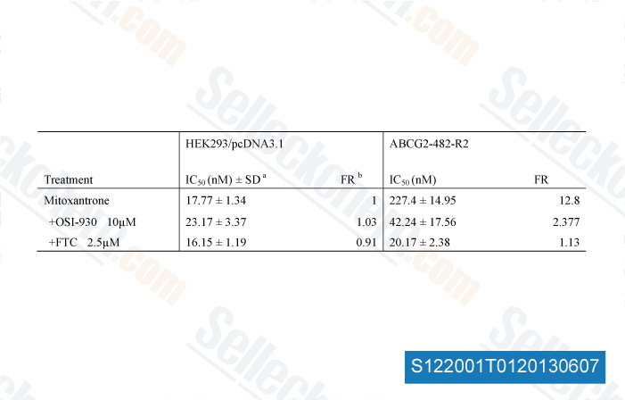
OSI-930 analogues as novel reversal agents for ABCG2-mediated multidrug resistance. [Kuang Y, et al. Biochem Pharmacol, 2012, 84(6):766-74] PubMed: 22750060

RÜCKGABERICHTLINIE
Die bedingungslose Rückgaberichtlinie von Selleck Chemical gewährleistet unseren Kunden ein reibungsloses Online-Einkaufserlebnis. Wenn Sie in irgendeiner Weise mit Ihrem Kauf unzufrieden sind, können Sie jeden Artikel innerhalb von 7 Tagen nach Erhalt zurückgeben. Im Falle von Produktqualitätsproblemen, sei es protokollbezogene oder produktbezogene Probleme, können Sie jeden Artikel innerhalb von 365 Tagen ab dem ursprünglichen Kaufdatum zurückgeben. Bitte befolgen Sie die nachstehenden Anweisungen, wenn Sie Produkte zurücksenden.

VERSAND UND LAGERUNG
Selleck-Produkte werden bei Raumtemperatur transportiert. Wenn Sie das Produkt bei Raumtemperatur erhalten, seien Sie versichert, dass die Qualitätskontrollabteilung von Selleck Experimente durchgeführt hat, um zu überprüfen, dass die normale Temperaturplatzierung von einem Monat die biologische Aktivität von Pulverprodukten nicht beeinträchtigt. Nach dem Sammeln lagern Sie das Produkt bitte gemäß den in der Datenblatt beschriebenen Anforderungen. Die meisten Selleck-Produkte sind unter den empfohlenen Bedingungen stabil.

NICHT FÜR DIE ANWENDUNG AM MENSCHEN, FÜR VETERINÄRMEDIZINISCHE DIAGNOSTIK ODER THERAPEUTISCHE ZWECKE.