Technische Daten
| Formel | C32H34Cl2N4O4S |
||||||
| Molekulargewicht | 641.61 | CAS-Nr. | 477575-56-7 | ||||
| Löslichkeit (25°C)* | In vitro | DMSO | 100 mg/mL (155.85 mM) | ||||
| Water | Insoluble | ||||||
| Ethanol | Insoluble | ||||||
| In vivo (Lösungsmittel einzeln und der Reihe nach zum Produkt hinzufügen.) |
|
||||||
|
* <1 mg/ml bedeutet schwer löslich oder unlöslich. * Bitte beachten Sie, dass Selleck die Löslichkeit aller Verbindungen intern testet und die tatsächliche Löslichkeit geringfügig von veröffentlichten Werten abweichen kann. Dies ist normal und ist auf geringfügige Batch-zu-Batch-Variationen zurückzuführen. * Versand bei Raumtemperatur (Stabilitätstests zeigen, dass dieses Produkt ohne Kühlmaßnahmen versendet werden kann.) |
|||||||
Vorbereitung von Stammlösungen
Biologische Aktivität
| Beschreibung | PHA-665752 ist ein potenter, selektiver und ATP-kompetitiver c-Met Inhibitor mit einer IC50 von 9 nM in zellfreien Assays, mit einer >50-fachen Selektivität für c-Met gegenüber RTKs oder STKs. | ||||||
|---|---|---|---|---|---|---|---|
| Ziele |
|
||||||
| In vitro | PHA-665752 hemmt die c-Met-Kinaseaktivität signifikant mit einem Ki von 4 nM und zeigt eine >50-fache Selektivität für c-Met im Vergleich zu verschiedenen Tyrosin- und Serin-Threonin-Kinasen. Diese Verbindung hemmt die HGF-stimulierte c-Met-Autophosphorylierung potent mit einer IC50 von 25-50 nM. Sie blockiert auch signifikant HGF- und c-Met-abhängige Funktionen wie Zellmotilität und Zellproliferation mit IC50-Werten von 40-50 nM bzw. 18-42 nM. Darüber hinaus hemmt diese Chemikalie die HGF-stimulierte oder konstitutive Phosphorylierung von Mediatoren downstream von c-Met wie Gab-1, ERK, Akt, STAT3, PLC-γ und FAK in mehreren Tumorzelllinien potent. Es hemmt das Zellwachstum in TPR-MET-transformierten BaF3-Zellen mit einer IC50 von <60 nM und hemmt die konstitutive Zellmotilität und -migration um 92,5 % bei 0,2 μM. Die Hemmung von c-Met durch diese Verbindung (0,2 μM) induziert auch eine Zellapoptose von 33,1 % und einen G1-Zellzyklusarrest, wobei die Zellen in der G1-Phase von 42,4 % auf 77,0 % ansteigen. Es kann mit Rapamycin zusammenwirken, um das Zellwachstum von TPR-MET-transformierten BaF3-Zellen und nicht-kleinzelligen Lungenkrebs-H441-Zellen zu hemmen. | ||||||
| In vivo | Die Verabreichung von PHA-665752 induziert eine dosisabhängige Tumorwachstumshemmung von S114-Xenotransplantaten um 20 %, 39 % und 68 % bei Dosen von 7,5, 15 bzw. 30 mg/kg/Tag. Diese Verbindung reduzierte das Tumorwachstum von NCI-H69, NCI-H441 und A549 in Maus-Xenotransplantaten signifikant um 99 %, 75 % bzw. 59 %. Sie hemmt auch die Angiogenese signifikant um >85 %, da sie die Produktion des vaskulären endothelialen Wachstumsfaktors verringert und die Produktion des Angiogenese-Inhibitors Thrombospondin-1 erhöht. |
Protokoll (aus Referenz)
| Kinase-Assay:[1] |
|
|---|---|
| Zell-Assay:[1] |
|
| Tierstudie:[1] |
|
Referenzen
|
Kundenproduktvalidierung
-
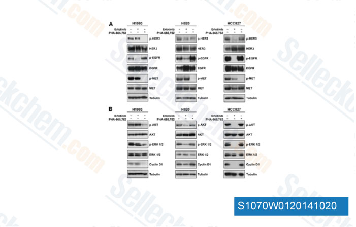
Daten von [ Clin Cancer Res , 2014 , 20, 4806-15 ]
-
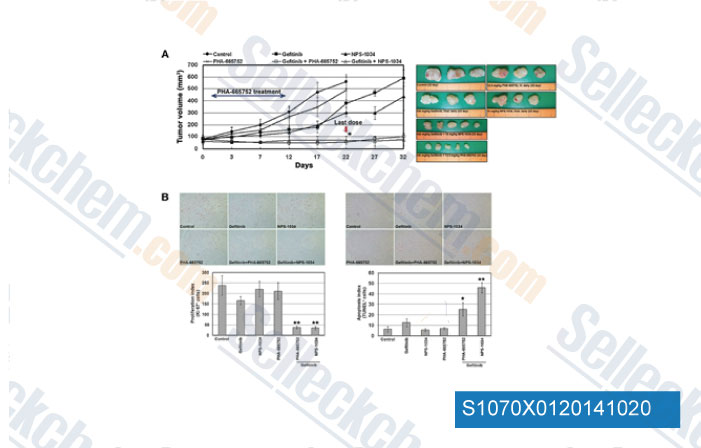
Daten von [ Cancer Res , 2014 , 74, 253-62 ]
-
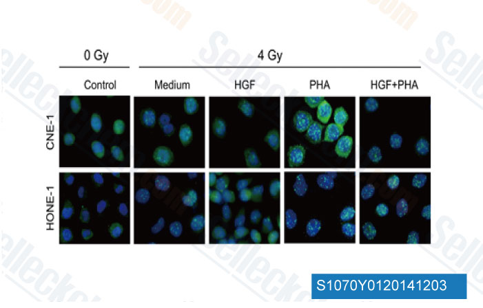
Daten von [ Biochem Biophys Res Commun , 2014 , 449(1), 49-54 ]
-
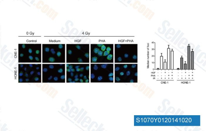
Daten von [ Biochem Biophys Res Commun , 2014 , 449, 49-54 ]
Sellecks PHA-665752 Wurde zitiert von 100 Publikationen
| Inhibition of TFF3 synergizes with c-MET inhibitors to decrease the CSC-like phenotype and metastatic burden in ER+HER2+ mammary carcinoma [ Cell Death Dis, 2025, 16(1):76] | PubMed: 39920140 |
| Macrophage Subpopulation Promotes Skeletal Muscle Regeneration Through HGF/MET Signaling-Mediated Skeletal Muscle Stem Cell Proliferation [ Aging Cell, 2025, e70042.] | PubMed: 40132988 |
| Overcoming MET-targeted drug resistance in MET-amplified lung cancer by aurora kinase B inhibition [ Biochim Biophys Acta Mol Cell Res, 2025, 1872(7):120001] | PubMed: 40499687 |
| MET receptor serves as a promising target in melanoma brain metastases [ Acta Neuropathol, 2024, 147(1):44] | PubMed: 38386085 |
| LncRNA CHROMR/miR-27b-3p/MET axis promotes the proliferation, invasion, and contributes to rituximab resistance in diffuse large B-cell lymphoma [ J Biol Chem, 2024, 300(3):105762] | PubMed: 38367665 |
| Single targeting of MET in EGFR-mutated and MET-amplified non-small cell lung cancer [ Br J Cancer, 2023, 128(12):2186-2196] | PubMed: 37059804 |
| Cancer-Associated Fibroblasts Promote Radioresistance of Breast Cancer Cells via the HGF/c-Met Signaling Pathway [ Int J Radiat Oncol Biol Phys, 2023, 116(3):640-654] | PubMed: 36586496 |
| MET Receptor Tyrosine Kinase Inhibition Reduces Interferon-Gamma (IFN-γ)-Stimulated PD-L1 Expression through the STAT3 Pathway in Melanoma Cells [ Cancers (Basel), 2023, 15(13)3408] | PubMed: 37444518 |
| Cabozantinib inhibits HBV-RNA transcription by decreasing STAT3 binding to the enhancer region of cccDNA [ Hepatol Commun, 2023, 10.1097/HC9.0000000000000313] | PubMed: 37938099 |
| Dual-targeting therapy against HER3/MET in human colorectal cancers [ Cancer Med, 2023, 10.1002/cam4.5673] | PubMed: 36751113 |
RÜCKGABERICHTLINIE
Die bedingungslose Rückgaberichtlinie von Selleck Chemical gewährleistet unseren Kunden ein reibungsloses Online-Einkaufserlebnis. Wenn Sie in irgendeiner Weise mit Ihrem Kauf unzufrieden sind, können Sie jeden Artikel innerhalb von 7 Tagen nach Erhalt zurückgeben. Im Falle von Produktqualitätsproblemen, sei es protokollbezogene oder produktbezogene Probleme, können Sie jeden Artikel innerhalb von 365 Tagen ab dem ursprünglichen Kaufdatum zurückgeben. Bitte befolgen Sie die nachstehenden Anweisungen, wenn Sie Produkte zurücksenden.
VERSAND UND LAGERUNG
Selleck-Produkte werden bei Raumtemperatur transportiert. Wenn Sie das Produkt bei Raumtemperatur erhalten, seien Sie versichert, dass die Qualitätskontrollabteilung von Selleck Experimente durchgeführt hat, um zu überprüfen, dass die normale Temperaturplatzierung von einem Monat die biologische Aktivität von Pulverprodukten nicht beeinträchtigt. Nach dem Sammeln lagern Sie das Produkt bitte gemäß den in der Datenblatt beschriebenen Anforderungen. Die meisten Selleck-Produkte sind unter den empfohlenen Bedingungen stabil.
NICHT FÜR DIE ANWENDUNG AM MENSCHEN, FÜR VETERINÄRMEDIZINISCHE DIAGNOSTIK ODER THERAPEUTISCHE ZWECKE.