Technische Daten
| Formel | C14H13BrN2O |
||||||||||
| Molekulargewicht | 305.17 | CAS-Nr. | 65678-07-1 | ||||||||
| Löslichkeit (25°C)* | In vitro | DMSO | 61 mg/mL (199.88 mM) | ||||||||
| Water | Insoluble | ||||||||||
| Ethanol | Insoluble | ||||||||||
| In vivo (Lösungsmittel einzeln und der Reihe nach zum Produkt hinzufügen.) |
|
||||||||||
|
* <1 mg/ml bedeutet schwer löslich oder unlöslich. * Bitte beachten Sie, dass Selleck die Löslichkeit aller Verbindungen intern testet und die tatsächliche Löslichkeit geringfügig von veröffentlichten Werten abweichen kann. Dies ist normal und ist auf geringfügige Batch-zu-Batch-Variationen zurückzuführen. * Versand bei Raumtemperatur (Stabilitätstests zeigen, dass dieses Produkt ohne Kühlmaßnahmen versendet werden kann.) |
|||||||||||
Vorbereitung von Stammlösungen
Biologische Aktivität
| Beschreibung | AG-1024 (Tyrphostin, AGS 200) hemmt die IGF-1R-Autophosphorylierung mit einem IC50 von 7 μM, ist bei IR mit einem IC50 von 57 μM weniger potent und unterscheidet spezifisch zwischen InsR und IGF-1R (im Vergleich zu anderen Tyrphostinen). | ||||
|---|---|---|---|---|---|
| Ziele |
|
||||
| In vitro | AG-1024 blockiert die IGF-1R- und IR-Autophosphorylierung mit einer IC50 von 7 μM bzw. 57 μM. Diese Verbindung hemmt auch die Protein Tyrosine Kinase-Aktivität gegenüber exogenen Substraten (TKA) mit IC50-Werten von 18 μM bzw. 80 μM. Diese Chemikalie (10 μM) hemmt die Zellproliferation zeitabhängig und induziert die Apoptose in MCF-7-Zellen nach 48 Stunden um 20,1 % und um >40 %, wenn sie mit Bestrahlung (10 Gy) kombiniert wird, was potenter ist als die alleinige Bestrahlung (10 Gy) um 11,8 %, was mit einer Herunterregulierung von Phospho-Akt1 und Bcl-2 sowie einer Hochregulierung von Bax, p53 und p21 assoziiert ist. Es hemmt signifikant die Proliferation von Melanomzellen mit einer IC50 von <50 nM in Abwesenheit von Serum, indem es die MAPK/ERK2-Signalübertragung blockiert, anschließend schnell die pRb-Dephosphorylierung und -Aktivierung induziert und schließlich die Bildung von wachstumshemmenden pRb-E2F-Komplexen. Die Behandlung mit dieser Verbindung reguliert die Expression von Bcr-Abl und P-Akt herunter und die DNA-PKcs-Expression in UT7-9- und Ba/F3-p210-Zellen hoch, was zu einer verminderten klonogenen Überlebensrate und Proliferation führt. Es hemmt auch signifikant die Proliferation von Zellen, die gegen den BCR-ABL-Inhibitor STI571 resistent sind, was mit einer dosisabhängigen Abnahme der Bcr-Abl-Proteinexpression korreliert. | ||||
| In vivo | Die Verabreichung von AG-1024 in einer Dosis von 30 μg über 10 Tage hemmt signifikant das Tumorwachstum von Ba/F3-p210-Xenotransplantaten in Mäusen. |
Protokoll (aus Referenz)
| Kinase-Assay:[1] |
|
|---|---|
| Zell-Assay:[2] |
|
| Tierstudie:[4] |
|
Referenzen
|
Kundenproduktvalidierung
-
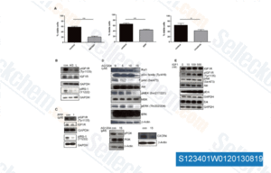
Daten von [ Blood , 2013 , 122(9), 1621-33 ]
-
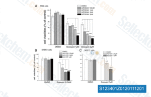
Daten von [ Mol Endocrinol , 2011 , 25, 2041-53 ]
-
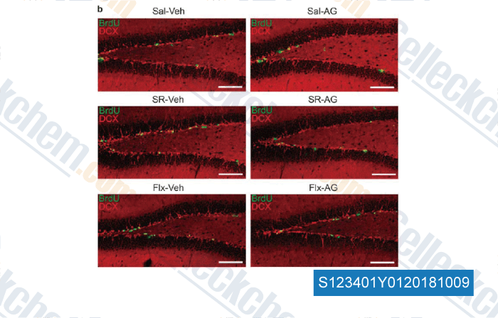
Daten von [ , , Mol Psychiatry, 2018, 23(4):833-842 ]
Sellecks AG-1024 Wurde zitiert von 27 Publikationen
| A Transcriptomics Analysis of the Regulation of Lens Fiber Cell Differentiation in the Absence of FGFRs and PTEN [ Cells, 2024, 13(14)1222] | PubMed: 39056803 |
| LTK and ALK promote neuronal polarity and cortical migration by inhibiting IGF1R activity [ EMBO Rep, 2023, 24(7):e56937] | PubMed: 37291945 |
| Ras-mutant cancers are sensitive to small molecule inhibition of V-type ATPases in mice [ Nat Biotechnol, 2022, 10.1038/s41587-022-01386-z] | PubMed: 35879364 |
| Tyrphostin AG1024 Suppresses Coronaviral Replication by Downregulating JAK1 via an IR/IGF-1R Independent Proteolysis Mediated by Ndfip1/2_NEDD4-like E3 Ligase Itch [ Pharmaceuticals (Basel), 2022, 15(2)241] | PubMed: 35215353 |
| Targeting oncogenic mutations in colorectal cancer using cryptotanshinone [ PLoS One, 2021, 16(2):e0247190] | PubMed: 33596259 |
| Regulation of Hippo-YAP Signaling by Insulin-Like Growth factor-1 Receptor in the Tumorigenesis of Diffuse Large B-cell Lymphoma [ J Hematol Oncol, 2020, 13(1):77] | PubMed: 32546241 |
| Myeloid cells provide critical support for T-ALL in vivo [ The University of Texas at Austin, 2020, ] | PubMed: None |
| An Autocrine IL-6/IGF-1R Loop Mediates EMT and Promotes Tumor Growth in Non-small Cell Lung Cancer. [ Int J Biol Sci, 2019, 15(9):1882-1891] | PubMed: 31523190 |
| mTORC2-mediated PDHE1α nuclear translocation links EBV-LMP1 reprogrammed glucose metabolism to cancer metastasis in nasopharyngeal carcinoma. [ Oncogene, 2019, 38(24):4669-4684] | PubMed: 30745576 |
| A novel 5HT3 receptor–IGF1 mechanism distinct from SSRI-induced antidepressant effects [Kondo M, et al. Mol Psychiatry, 2018, 23(4):833-842] | PubMed: 28439104 |
RÜCKGABERICHTLINIE
Die bedingungslose Rückgaberichtlinie von Selleck Chemical gewährleistet unseren Kunden ein reibungsloses Online-Einkaufserlebnis. Wenn Sie in irgendeiner Weise mit Ihrem Kauf unzufrieden sind, können Sie jeden Artikel innerhalb von 7 Tagen nach Erhalt zurückgeben. Im Falle von Produktqualitätsproblemen, sei es protokollbezogene oder produktbezogene Probleme, können Sie jeden Artikel innerhalb von 365 Tagen ab dem ursprünglichen Kaufdatum zurückgeben. Bitte befolgen Sie die nachstehenden Anweisungen, wenn Sie Produkte zurücksenden.
VERSAND UND LAGERUNG
Selleck-Produkte werden bei Raumtemperatur transportiert. Wenn Sie das Produkt bei Raumtemperatur erhalten, seien Sie versichert, dass die Qualitätskontrollabteilung von Selleck Experimente durchgeführt hat, um zu überprüfen, dass die normale Temperaturplatzierung von einem Monat die biologische Aktivität von Pulverprodukten nicht beeinträchtigt. Nach dem Sammeln lagern Sie das Produkt bitte gemäß den in der Datenblatt beschriebenen Anforderungen. Die meisten Selleck-Produkte sind unter den empfohlenen Bedingungen stabil.
NICHT FÜR DIE ANWENDUNG AM MENSCHEN, FÜR VETERINÄRMEDIZINISCHE DIAGNOSTIK ODER THERAPEUTISCHE ZWECKE.