|
Wie zu zitieren 1. Für Zitate im Text (Materialien & Methoden): 2. Für die Tabelle der Schlüsselressourcen: |
||
|
Gebührenfrei: (877) 796-6397 -- Nur USA und Kanada -- |
Fax: +1-832-582-8590 Bestellungen: +1-832-582-8158 |
Technischer Support: +1-832-582-8158 Ext:3 Bitte geben Sie Ihre Bestellnummer in der E-Mail an. Wir bemühen uns, alle E-Mail-Anfragen innerhalb eines Werktages zu beantworten. |
Biologische Beschreibung
| Spezifität | Akt1 Antibody [C19L11] erkennt endogene Spiegel des gesamten Akt1-Proteins. |
|---|---|
| Hintergrund | AKT, ein zentrales Protein in vielen zellulären Signalwegen, spielt eine entscheidende Rolle beim Zellüberleben, der Proliferation, der Glukoseaufnahme, dem Stoffwechsel, der Angiogenese und den Reaktionen auf Bestrahlung und Medikamente. Es gibt drei Isoformen von AKT: AKT1, AKT2 und AKT3, jede mit unterschiedlichen physiologischen Funktionen, Eigenschaften und Expressionsmustern, die je nach Zelltyp variieren. AKT1, auch bekannt als v-akt murine thymoma virales Onkogen-Homolog 1, ist eine Serin/Threonin-Proteinkinase. Ihre Aktivierung erfolgt durch Phosphorylierung an Serin 473 und Threonin 308. AKT1 ist hauptsächlich für die Phosphorylierung und Lokalisierung von CKDN1A (Cyclin-abhängiger Kinase-Inhibitor 1, auch bekannt als p21) verantwortlich. Interessanterweise wird p21, ein CDK-Inhibitor, der normalerweise im Zellkern vorkommt, oft von AKT1 ins Zytoplasma verlagert. Diese zytoplasmatische Lokalisierung von p21, die durch AKT1 ermöglicht wird, ist bei verschiedenen Krebsarten häufig, wo sie die Tumorentstehung durch Hemmung von für die Apoptose essentiellen Proteinen fördert. Eine erhöhte Expression verschiedener AKT-Isoformen wurde bei mehreren Krebsarten beobachtet. Insbesondere wurden erhöhte AKT1-Spiegel mit gastrointestinalen Stromatumoren (GIST), Schilddrüsenkrebs und Brustkrebs in Verbindung gebracht und dienen als Indikatoren für eine schlechte Prognose. |
Nutzungsinformationen
| Anwendung | WB, IP | Verdünnung |
|
||||
|---|---|---|---|---|---|---|---|
| Reaktivität | Human, Mouse, Rat, Monkey | ||||||
| Quelle | Rabbit Monoclonal Antibody | MW | 60 kDa | ||||
| Lagerpuffer | PBS, pH 7.2+50% Glycerol+0.05% BSA+0.01% NaN₃ | Lagerung (Ab dem Datum des Erhalts) |
–20°C (avoid freeze-thaw cycles), 2 years | ||||
| WB |
Experimental Protocol:
Sample preparation
1. Tissue: Lyse the tissue sample by adding an appropriate volume of ice-cold RIPA/NP-40 Lysis Buffer (containing Protease Inhibitor Cocktail),and homogenize the tissue at a low temperature or lyse it by sonication on ice, then incubate on ice for 30 minutes. 2. Adherent cell: Aspirate the culture medium and transfer the cells into an EP tube. Wash the cells with ice-cold PBS twice. Add an appropriate volume of RIPA/NP-40 Lysis Buffer (containing Protease Inhibitor Cocktail), sonicate to lyse the cells, and incubate on ice for 30 minutes. 3. Suspension cell: Transfer the culture medium to a pre-cooled centrifuge tube. Centrifuge and aspirate the supernatant. Wash the cells with ice-cold PBS twice.Add an appropriate volume of RIPA/NP-40 Lysis Buffer (containing Protease Inhibitor Cocktail), sonicate to lyse the cells, and incubate on ice for 30 minutes. 4. Place the lysate into a pre-cooled microcentrifuge tube. Centrifuge at 4°C for 15 min. Collect the supernatant;
5. Remove a small volume of lysate to determine the protein concentration;
6. Combine the lysate with protein loading buffer. Boil 20 µL sample under 95-100°C for 5 min. Centrifuge for 5 min after cool down on ice.
Electrophoretic separation
1. According to the concentration of extracted protein, load appropriate amount of protein sample and marker onto SDS-PAGE gels for electrophoresis. Recommended separating gel (lower gel) concentration: 10%. Reference Table for Selecting SDS-PAGE Separation Gel Concentrations 2. Power up 80V for 30 minutes. Then the power supply is adjusted (110 V~150 V), the Marker is observed, and the electrophoresis can be stopped when the indicator band of the predyed protein Marker where the protein is located is properly separated. (Note that the current should not be too large when electrophoresis, too large current (more than 150 mA) will cause the temperature to rise, affecting the result of running glue. If high currents cannot be avoided, an ice bath can be used to cool the bath.)
Transfer membrane
1. Take out the converter, soak the clip and consumables in the pre-cooled converter;
2. Activate PVDF membrane with methanol for 1 min and rinse with transfer buffer;
3. Install it in the order of "black edge of clip - sponge - filter paper - filter paper - glue -PVDF membrane - filter paper - filter paper - sponge - white edge of clip"; 4. The protein was electrotransferred to PVDF membrane. ( 0.45 µm PVDF membrane is recommended ) Reference Table for Selecting PVDF Membrane Pore Size Specifications Recommended conditions for wet transfer: 200 mA, 120 min. ( Note that the transfer conditions can be adjusted according to the protein size. For high-molecular-weight proteins, a higher current and longer transfer time are recommended. However, ensure that the transfer tank remains at a low temperature to prevent gel melting.)
Block
1. After electrotransfer, wash the film with TBST at room temperature for 5 minutes;
2. Incubate the film in the blocking solution for 1 hour at room temperature;
3. Wash the film with TBST for 3 times, 5 minutes each time.
Antibody incubation
1. Use 5% skim milk powder to prepare the primary antibody working liquid (recommended dilution ratio for primary antibody 1:1000), gently shake and incubate with the film at 4°C overnight; 2. Wash the film with TBST 3 times, 5 minutes each time;
3. Add the secondary antibody to the blocking solution and incubate with the film gently at room temperature for 1 hour;
4. After incubation, wash the film with TBST 3 times for 5 minutes each time.
Antibody staining
37. Add the prepared ECL luminescent substrate (or select other color developing substrate according to the second antibody) and mix evenly;
2. Incubate with the film for 1 minute, remove excess substrate (keep the film moist), wrap with plastic film, and expose in the imaging system.
|
Referenzen
|
Anwendungsdaten
WB
Validiert von Selleck
-
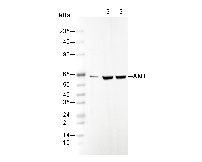 Lane 1: HeLA
Lane 2: NIH/3T3
Lane 3: C6