|
Wie zu zitieren 1. Für Zitate im Text (Materialien & Methoden): 2. Für die Tabelle der Schlüsselressourcen: |
||
|
Gebührenfrei: (877) 796-6397 -- Nur USA und Kanada -- |
Fax: +1-832-582-8590 Bestellungen: +1-832-582-8158 |
Technischer Support: +1-832-582-8158 Ext:3 Bitte geben Sie Ihre Bestellnummer in der E-Mail an. Wir bemühen uns, alle E-Mail-Anfragen innerhalb eines Werktages zu beantworten. |
Biologische Beschreibung
| Spezifität | BATF Antibody [G1B2] weist endogene Spiegel des gesamten BATF-Proteins nach. |
|---|---|
| Hintergrund | BATF (Basic Leucine Zipper ATF-like Transcription Factor) ist ein zentraler Transkriptionsfaktor in der AP-1-Familie, der die Differenzierung und Funktion von Immunzellen steuert. Es besitzt eine basische Leucin-Zipper (bZIP)-Domäne, die die Dimerisierung mit Jun- und Fos-Proteinen und eine spezifische DNA-Bindung ermöglicht, obwohl es keine Transaktivierungsdomäne besitzt und auf die Interaktion mit Kofaktoren zur Transkriptionsregulation angewiesen ist. BATF steuert Genexpressionsprogramme, die für die Entwicklung und Effektorfunktionen von T-Zellen (einschließlich Th17, Tfh und CD8+ zytolytische T-Zellen) und B-Zellen entscheidend sind, indem es Heterodimere bildet, die an TRE/CRE-Stellen binden und mit Jun/Fos-Dimeren konkurrieren, um die Transkription fein zu regulieren. BATF kooperiert mit Transkriptionsfaktoren wie IRF4 und STAT3 und orchestriert epigenomische und transkriptomische Umgestaltung, um Immunantworten zu fördern. Es unterdrückt direkt Sirt1, eine Histon-Deacetylase, wodurch die Histonacetylierung verbessert und Gene wie T-bet, die für die T-Zelldifferenzierung und das Überleben essentiell sind, aktiviert werden. BATF moduliert auch den Zellstoffwechsel, indem es die NAD+- und ATP-Produktion erhöht, um den Energiebedarf während der Immunaktivierung zu decken. Die Dysregulation von BATF führt zu Immunstörungen, Entzündungen und Krebs. |
Nutzungsinformationen
| Anwendung | WB, IP, FCM | Verdünnung |
|
||||||
|---|---|---|---|---|---|---|---|---|---|
| Reaktivität | Human, Mouse | ||||||||
| Quelle | Rabbit Monoclonal Antibody | MW | 15 kDa | ||||||
| Lagerpuffer | PBS, pH 7.2+50% Glycerol+0.05% BSA+0.01% NaN3 | Lagerung (Ab dem Datum des Erhalts) |
-20°C (avoid freeze-thaw cycles), 2 years | ||||||
| WB |
Experimental Protocol:
Sample preparation
1. Tissue: Lyse the tissue sample by adding an appropriate volume of ice-cold RIPA/NP-40 Lysis Buffer (containing Protease Inhibitor Cocktail),and homogenize the tissue at a low temperature. 2. Adherent cell: Aspirate the culture medium and wash the cells with ice-cold PBS twice. Lyse the cells by adding an appropriate volume of RIPA/NP-40 Lysis Buffer (containing Protease Inhibitor Cocktail) and put the sample on ice for 5 min. 3. Suspension cell: Transfer the culture medium to a pre-cooled centrifuge tube. Centrifuge and aspirate the supernatant. Wash the cells with ice-cold PBS twice. Lyse the cells by adding an appropriate volume of RIPA/NP-40 Lysis Buffer (containing Protease Inhibitor Cocktail) and put the sample on ice for 5 min. 4. Place the lysate into a pre-cooled microcentrifuge tube. Centrifuge at 4°C for 15 min. Collect the supernatant;
5. Remove a small volume of lysate to determine the protein concentration;
6. Combine the lysate with protein loading buffer. Boil 20 µL sample under 95-100°C for 5 min. Centrifuge for 5 min after cool down on ice.
Electrophoretic separation
1. According to the concentration of extracted protein, load appropriate amount of protein sample and marker onto SDS-PAGE gels for electrophoresis. Recommended separating gel (lower gel) concentration: 20%. Reference Table for Selecting SDS-PAGE Separation Gel Concentrations 2. Power up 80V for 30 minutes. Then the power supply is adjusted (110 V~150 V), the Marker is observed, and the electrophoresis can be stopped when the indicator band of the predyed protein Marker where the protein is located is properly separated. (Note that the current should not be too large when electrophoresis, too large current (more than 150 mA) will cause the temperature to rise, affecting the result of running glue. If high currents cannot be avoided, an ice bath can be used to cool the bath.)
Transfer membrane
1. Take out the converter, soak the clip and consumables in the pre-cooled converter;
2. Activate PVDF membrane with methanol for 1 min and rinse with transfer buffer;
3. Install it in the order of "black edge of clip - sponge - filter paper - filter paper - glue -PVDF membrane - filter paper - filter paper - sponge - white edge of clip"; 4. The protein was electrotransferred to PVDF membrane. ( 0.22 µm PVDF membrane is recommended )) Reference Table for Selecting PVDF Membrane Pore Size Specifications Recommended conditions for wet transfer: 200 mA, 60 min. ( Note that the transfer conditions can be adjusted according to the protein size. For high-molecular-weight proteins, a higher current and longer transfer time are recommended. However, ensure that the transfer tank remains at a low temperature to prevent gel melting.)
Block
1. After electrotransfer, wash the film with TBST at room temperature for 5 minutes;
2. Incubate the film in the blocking solution for 1 hour at room temperature;
3. Wash the film with TBST for 3 times, 5 minutes each time.
Antibody incubation
1. Use 5% skim milk powder to prepare the primary antibody working liquid (recommended dilution ratio for primary antibody 1:1000), gently shake and incubate with the film at 4°C overnight; 2. Wash the film with TBST 3 times, 5 minutes each time;
3. Add the secondary antibody to the blocking solution and incubate with the film gently at room temperature for 1 hour;
4. After incubation, wash the film with TBST 3 times for 5 minutes each time.
Antibody staining
1. Add the prepared ECL luminescent substrate (or select other color developing substrate according to the second antibody) and mix evenly;
2. Incubate with the film for 1 minute, remove excess substrate (keep the film moist), wrap with plastic film, and expose in the imaging system. (Exposure time of at least 60s is recommended)
|
Referenzen
|
Anwendungsdaten
WB
Validiert von Selleck
-
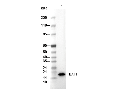 Lane 1: HDLM-2