|
Wie zu zitieren 1. Für Zitate im Text (Materialien & Methoden): 2. Für die Tabelle der Schlüsselressourcen: |
||
|
Gebührenfrei: (877) 796-6397 -- Nur USA und Kanada -- |
Fax: +1-832-582-8590 Bestellungen: +1-832-582-8158 |
Technischer Support: +1-832-582-8158 Ext:3 Bitte geben Sie Ihre Bestellnummer in der E-Mail an. Wir bemühen uns, alle E-Mail-Anfragen innerhalb eines Werktages zu beantworten. |
Biologische Beschreibung
| Spezifität | β Amyloid (Aβ) 1-42 Antibody [H15J22] detektiert endogene Spiegel des gesamten β Amyloid 1-42 Proteins. |
|---|---|
| Hintergrund | Amyloid β 1-42 (Aβ42) ist ein Peptid aus 42 Aminosäuren, das aus dem Amyloid-Vorläuferprotein (APP) durch sequentielle Spaltung durch β-Sekretase und γ-Sekretase in Neuronen entsteht. Strukturell ist Aβ42 an seinem C-Terminus stark hydrophob, was eine Konformationsverschiebung von α-Helix zu β-Faltblatt fördert, die die Aggregation zu löslichen Oligomeren, Protofibrillen und unlöslichen Amyloidfibrillen erleichtert. Aβ42 wird hauptsächlich im Gehirn, insbesondere im Neokortex, exprimiert und ist aggregationsanfälliger und neurotoxischer als die kürzere Aβ40-Form. Funktionell ist die pathologische Akkumulation von Aβ42 – insbesondere als lösliche Oligomere – stark an der Pathogenese der Alzheimer-Krankheit beteiligt, indem sie die synaptische Signalübertragung stört, das Gedächtnis beeinträchtigt und zur Plaquebildung beiträgt. |
Nutzungsinformationen
| Anwendung | IHC | Verdünnung |
|
||
|---|---|---|---|---|---|
| Reaktivität | Mouse, Rat, Human | ||||
| Quelle | Rabbit Monoclonal Antibody | MW | |||
| Lagerpuffer | PBS, pH 7.2+50% Glycerol+0.05% BSA+0.01% NaN3 | Lagerung (Ab dem Datum des Erhalts) |
-20°C (avoid freeze-thaw cycles), 2 years | ||
| IHC |
Experimental Protocol:
Deparaffinization/Rehydration
1. Deparaffinize/hydrate sections:
2. Incubate sections in three washes of xylene for 5 min each.
3. Incubate sections in two washes of 100% ethanol for 10 min each.
4. Incubate sections in two washes of 95% ethanol for 10 min each.
5. Wash sections two times in dH2O for 5 min each.
6.Antigen retrieval: For Citrate: Heat slides in a microwave submersed in 1X citrate unmasking solution until boiling is initiated; continue with 10 min at a sub-boiling temperature (95°-98°C). Cool slides on bench top for 30 min.
Staining
1. Wash sections in dH2O three times for 5 min each.
2. Incubate sections in 3% hydrogen peroxide for 10 min.
3. Wash sections in dH2O two times for 5 min each.
4. Wash sections in wash buffer for 5 min.
5. Block each section with 100–400 µl of blocking solution for 1 hr at room temperature.
6. Remove blocking solution and add 100–400 µl primary antibody diluent in to each section. Incubate overnight at 4°C.
7. Remove antibody solution and wash sections with wash buffer three times for 5 min each.
8. Cover section with 1–3 drops HRPas needed. Incubate in a humidified chamber for 30 min at room temperature.
9. Wash sections three times with wash buffer for 5 min each.
10. Add DAB Chromogen Concentrate to DAB Diluent and mix well before use.
11. Apply 100–400 µl DAB to each section and monitor closely. 1–10 min generally provides an acceptable staining intensity.
12. Immerse slides in dH2O.
13. If desired, counterstain sections with hematoxylin.
14. Wash sections in dH2O two times for 5 min each.
15. Dehydrate sections: Incubate sections in 95% ethanol two times for 10 sec each; Repeat in 100% ethanol, incubating sections two times for 10 sec each; Repeat in xylene, incubating sections two times for 10 sec each.
16. Mount sections with coverslips and mounting medium.
|
Referenzen
|
Anwendungsdaten
IHC
Validiert von Selleck
-
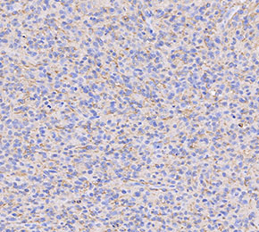 Immunohistochemical analysis of formalin fixed paraffin embedded human brain tissue with F3375 at 1:1000 dilution.