|
Wie zu zitieren 1. Für Zitate im Text (Materialien & Methoden): 2. Für die Tabelle der Schlüsselressourcen: |
||
|
Gebührenfrei: (877) 796-6397 -- Nur USA und Kanada -- |
Fax: +1-832-582-8590 Bestellungen: +1-832-582-8158 |
Technischer Support: +1-832-582-8158 Ext:3 Bitte geben Sie Ihre Bestellnummer in der E-Mail an. Wir bemühen uns, alle E-Mail-Anfragen innerhalb eines Werktages zu beantworten. |
Biologische Beschreibung
| Spezifität | CA19-9 Antibody [C17A13] detektiert endogene Mengen an CA 19-9-Protein. |
|---|---|
| Hintergrund | CA19-9 (Kohlenhydratantigen 19-9), auch bekannt als Sialyl Lewis A (sLeᵃ), ist ein sialyliertes Lewis-Blutgruppenantigen, ein Tetrasaccharid-Glykan-Epitop, das aus Sialinsäure, Galaktose, N-Acetylglucosamin und Fucose besteht und über den Lewis-Blutgruppenweg biosynthetisiert wird, wobei für seine Bildung ein funktionelles Lewis-Antigen erforderlich ist. CA19-9 wird auf Glykoproteinen und Glykolipiden von Epithelzellmembranen exprimiert, insbesondere in Pankreas, Gallenwegen, Magen und Dickdarm, und kann in den Blutkreislauf abgegeben werden. Es ist der aktuelle Goldstandard-Serum-Biomarker für Pankreasadenokarzinome. Funktionell dient CA19-9 als Biomarker (für Diagnose, Prognose, Überwachung des Therapieansprechens und Nachweis eines Rezidivs), als Prädiktor (korreliert mit Tumorlast, Stadium und Resektabilität) und als Promotor der Krebsrogression durch Erleichterung der E-Selectin-vermittelten Adhäsion, Verbesserung der Angiogenese, Modulation von Immunantworten und Beeinflussung von Tumor-Mikroumgebungs-Interaktionen. Neben Bauchspeicheldrüsenkrebs ist CA19-9 auch bei anderen gastrointestinalen Malignomen und bestimmten gutartigen Erkrankungen erhöht und wird als therapeutisches Ziel mittels Antikörpern, Impfstoffen, Biosynthese-Inhibitoren und CA19-9-gesteuerten Arzneimittelabgabesystemen untersucht. |
Nutzungsinformationen
| Anwendung | IHC, FCM, ELISA | Verdünnung |
|
||
|---|---|---|---|---|---|
| Reaktivität | Human | ||||
| Quelle | Mouse Monoclonal Antibody | MW | |||
| Lagerpuffer | PBS, pH 7.2+50% Glycerol+0.05% BSA+0.01% NaN3 | Lagerung (Ab dem Datum des Erhalts) |
-20°C (avoid freeze-thaw cycles), 2 years | ||
| IHC |
Experimental Protocol:
Deparaffinization/Rehydration
1. Deparaffinize/hydrate sections:
2. Incubate sections in three washes of xylene for 5 min each.
3. Incubate sections in two washes of 100% ethanol for 10 min each.
4. Incubate sections in two washes of 95% ethanol for 10 min each.
5. Wash sections two times in dH2O for 5 min each.
6.Antigen retrieval: For Citrate: Heat slides in a microwave submersed in 1X citrate unmasking solution until boiling is initiated; continue with 10 min at a sub-boiling temperature (95°-98°C). Cool slides on bench top for 30 min.
Staining
1. Wash sections in dH2O three times for 5 min each.
2. Incubate sections in 3% hydrogen peroxide for 10 min.
3. Wash sections in dH2O two times for 5 min each.
4. Wash sections in wash buffer for 5 min.
5. Block each section with 100–400 µl of blocking solution for 1 hr at room temperature.
6. Remove blocking solution and add 100–400 µl primary antibody diluent in to each section. Incubate overnight at 4°C.
7. Remove antibody solution and wash sections with wash buffer three times for 5 min each.
8. Cover section with 1–3 drops HRPas needed. Incubate in a humidified chamber for 30 min at room temperature.
9. Wash sections three times with wash buffer for 5 min each.
10. Add DAB Chromogen Concentrate to DAB Diluent and mix well before use.
11. Apply 100–400 µl DAB to each section and monitor closely. 1–10 min generally provides an acceptable staining intensity.
12. Immerse slides in dH2O.
13. If desired, counterstain sections with hematoxylin.
14. Wash sections in dH2O two times for 5 min each.
15. Dehydrate sections: Incubate sections in 95% ethanol two times for 10 sec each; Repeat in 100% ethanol, incubating sections two times for 10 sec each; Repeat in xylene, incubating sections two times for 10 sec each.
16. Mount sections with coverslips and mounting medium.
|
Referenzen
|
Anwendungsdaten
IHC
Validiert von Selleck
-
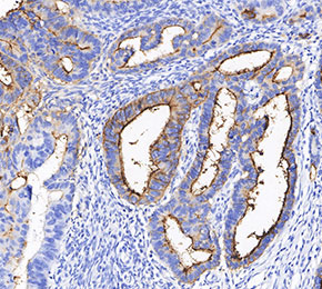 Immunohistochemical analysis of formalin fixed paraffin embedded human colorectal cancer tissue with F2466 at 1:100 dilution.