|
Wie zu zitieren 1. Für Zitate im Text (Materialien & Methoden): 2. Für die Tabelle der Schlüsselressourcen: |
||
|
Gebührenfrei: (877) 796-6397 -- Nur USA und Kanada -- |
Fax: +1-832-582-8590 Bestellungen: +1-832-582-8158 |
Technischer Support: +1-832-582-8158 Ext:3 Bitte geben Sie Ihre Bestellnummer in der E-Mail an. Wir bemühen uns, alle E-Mail-Anfragen innerhalb eines Werktages zu beantworten. |
Biologische Beschreibung
| Spezifität | CaV1.3 Antibody [G19D14] detektiert endogene Spiegel des gesamten CaV1.3-Proteins. |
|---|---|
| Hintergrund | Die kardiale Erregungs-Kontraktions-Kopplung bezieht sich auf die Abfolge von Ereignissen, bei denen die elektrische Stimulation eines Kardiomyozyten die Muskelkontraktion im Herzen auslöst. L-Typ-Ca²⁺-Kanäle sind für diesen Prozess von entscheidender Bedeutung, da sie den Kalziumeinstrom erleichtern und zur Membranerregbarkeit beitragen. Es wurden vier Subtypen von L-Typ-Ca²⁺-Kanälen identifiziert: Cav1.1, Cav1.2, Cav1.3 und Cav1.4. Cav1.1 findet sich überwiegend in der Skelettmuskulatur, während Cav1.4 hauptsächlich in der Netzhaut und bestimmten Immunzellen exprimiert wird. Cav1.3 ist im Herzen, in somatodendritischen Regionen von Neuronen, endokrinen Zellen und sensorischen Zellen vorhanden. Im Herzen wird die Cav1.3-Aktivität durch multiple Neurotransmitter moduliert. Die Phosphorylierung durch cAMP-abhängige Proteinkinase A (PKA) erfolgt an den Serinresten 1743 und 1816 innerhalb der C-terminalen Region. Proteinkinase C (PKC) reguliert Cav1.3 auch in einer Isozym-spezifischen Weise über die Phosphorylierung an Serin 81 in der N-terminalen Domäne. Zusätzlich beeinflusst alternatives Spleißen innerhalb des C-Terminus das Kanalverhalten, insbesondere die Abnahme der Ca²⁺-abhängigen Inaktivierung. Funktionell trägt Cav1.3 zur kardialen Schrittmacherfunktion und zur atrioventrikulären (AV-)Leitung bei. Eine Dysfunktion von Cav1.3 wurde mit Anomalien des Sinusknotens und des AV-Knotens sowie der Entwicklung von Vorhofflimmern in Verbindung gebracht. |
Nutzungsinformationen
| Anwendung | IHC, FCM | Verdünnung |
|
||
|---|---|---|---|---|---|
| Reaktivität | Human, Mouse | ||||
| Quelle | Mouse Monoclonal Antibody | MW | |||
| Lagerpuffer | PBS, pH 7.2+50% Glycerol+0.05% BSA+0.01% NaN3 | Lagerung (Ab dem Datum des Erhalts) |
-20°C (avoid freeze-thaw cycles), 2 years | ||
| IHC |
Experimental Protocol:
Deparaffinization/Rehydration
1. Deparaffinize/hydrate sections:
2. Incubate sections in three washes of xylene for 5 min each.
3. Incubate sections in two washes of 100% ethanol for 10 min each.
4. Incubate sections in two washes of 95% ethanol for 10 min each.
5. Wash sections two times in dH2O for 5 min each.
6.Antigen retrieval: For Citrate: Heat slides in a microwave submersed in 1X citrate unmasking solution until boiling is initiated; continue with 10 min at a sub-boiling temperature (95°-98°C). Cool slides on bench top for 30 min.
Staining
1. Wash sections in dH2O three times for 5 min each.
2. Incubate sections in 3% hydrogen peroxide for 10 min.
3. Wash sections in dH2O two times for 5 min each.
4. Wash sections in wash buffer for 5 min.
5. Block each section with 100–400 µl of blocking solution for 1 hr at room temperature.
6. Remove blocking solution and add 100–400 µl primary antibody diluent in to each section. Incubate overnight at 4°C.
7. Remove antibody solution and wash sections with wash buffer three times for 5 min each.
8. Cover section with 1–3 drops HRPas needed. Incubate in a humidified chamber for 30 min at room temperature.
9. Wash sections three times with wash buffer for 5 min each.
10. Add DAB Chromogen Concentrate to DAB Diluent and mix well before use.
11. Apply 100–400 µl DAB to each section and monitor closely. 1–10 min generally provides an acceptable staining intensity.
12. Immerse slides in dH2O.
13. If desired, counterstain sections with hematoxylin.
14. Wash sections in dH2O two times for 5 min each.
15. Dehydrate sections: Incubate sections in 95% ethanol two times for 10 sec each; Repeat in 100% ethanol, incubating sections two times for 10 sec each; Repeat in xylene, incubating sections two times for 10 sec each.
16. Mount sections with coverslips and mounting medium.
|
Referenzen
|
Anwendungsdaten
IHC
Validiert von Selleck
-
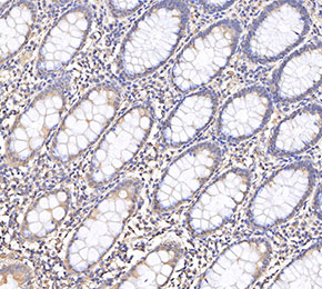 Immunohistochemical analysis of formalin fixed paraffin embedded human colorectal cancer tissue with F3798 at 1:1000 dilution.