Technische Daten
| Formel | C18H20ClN5 |
||||||||||
| Molekulargewicht | 341.84 | CAS-Nr. | 885499-61-6 | ||||||||
| Löslichkeit (25°C)* | In vitro | DMSO | 25 mg/mL (73.13 mM) | ||||||||
| Ethanol | 5 mg/mL (14.62 mM) | ||||||||||
| Water | Insoluble | ||||||||||
| In vivo (Lösungsmittel einzeln und der Reihe nach zum Produkt hinzufügen.) |
|
||||||||||
|
* <1 mg/ml bedeutet schwer löslich oder unlöslich. * Bitte beachten Sie, dass Selleck die Löslichkeit aller Verbindungen intern testet und die tatsächliche Löslichkeit geringfügig von veröffentlichten Werten abweichen kann. Dies ist normal und ist auf geringfügige Batch-zu-Batch-Variationen zurückzuführen. * Versand bei Raumtemperatur (Stabilitätstests zeigen, dass dieses Produkt ohne Kühlmaßnahmen versendet werden kann.) |
|||||||||||
Vorbereitung von Stammlösungen
Biologische Aktivität
| Beschreibung | CCT128930 ist ein potenter, ATP-kompetitiver und selektiver Inhibitor von Akt2 mit einem IC50 von 6 nM in einem zellfreien Assay, 28-fach höhere Selektivität für Akt2 als die eng verwandte PKA-Kinase. Diese Verbindung induziert Zellzyklusarrest, DNA-Schäden und Autophagy unabhängig von der Akt-Inhibition. Eine hohe Dosis dieser Chemikalie löst Zell-Apoptosis in HepG2-Zellen aus. | ||||||
|---|---|---|---|---|---|---|---|
| Ziele |
|
||||||
| In vitro | CCT128930 zeigt eine ausgeprägte antiproliferative Aktivität gegen PTEN-defiziente humane Tumorzelllinien, einschließlich U87MG-Glioblastomzellen, LNCaP-Prostatakrebszellen und PC3-Prostatakrebszellen, mit GI50-Werten von 6,3 μM, 0,35 μM bzw. 1,9 μM. Darüber hinaus verursacht diese Verbindung einen G1-Arrest in PTEN-null U87MG-Glioblastomzellen und eine Blockade des Akt-Signalwegs. | ||||||
| In vivo | CCT128930 bei 25 mg/kg i.p. zeigt einen ausgeprägten Antitumor-Effekt in etablierten PTEN-null U87MG-Glioblastom-Xenotransplantaten mit einem Behandlungs:Kontroll (T/C)-Verhältnis von 48% am Tag 12. In HER2-positiven, PIK3CA-mutierten BT474-Brustkrebs-Xenotransplantaten führt diese Verbindung bei 40 mg/kg ebenfalls zu einem tiefgreifenden Antitumor-Effekt mit vollständigem Wachstumsstillstand und einem T/C-Verhältnis von 29% am Tag 22. Diese chemische Substanz, intravenös verabreicht, erreicht eine Spitzenkonzentration von 6,4 μM im Plasma und wird mit einer relativ kurzen Halbwertszeit, einem hohen Verteilungsvolumen und einer schnellen Clearance eliminiert, was eine Fläche unter der Kurve AUC0-∞ von 4,6 μM·h ergibt. Intraperitoneal verabreicht, führt es zu einer Spitzen-Plasmakonzentration von 1,3 μM und der entsprechenden AUC0-∞ von 1,3 μM·h. Die orale Verabreichung dieser Verbindung führt zu einer Spitzen-Plasmakonzentration von nur 0,43 μM und einer entsprechend niedrigen AUC0-∞ von 0,4 μM·h. |
Protokoll (aus Referenz)
| Kinase-Assay:[1] |
|
|---|---|
| Zell-Assay:[1] |
|
| Tierstudie:[1] |
|
Referenzen
|
Kundenproduktvalidierung
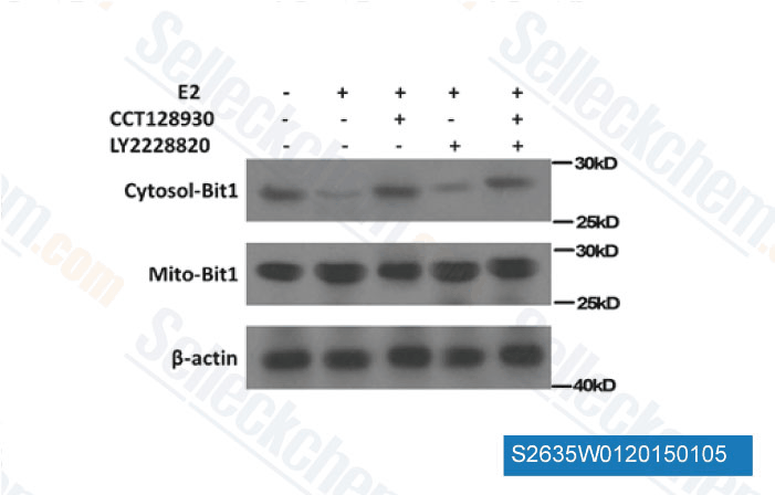
-
Daten von [ DNA Cell Biol , 2014 , 33(12), 847-53 ]
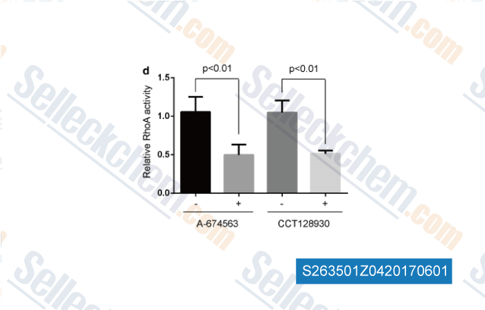
-
, , Cancer Cell Int, 2017, doi: 10.1186/s12935-017-0396-8
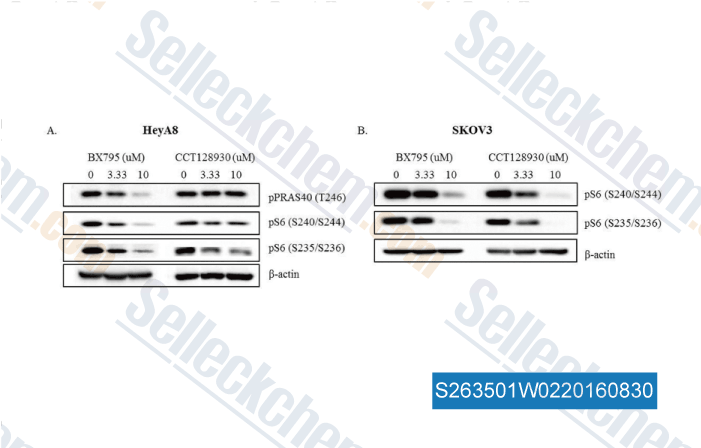
-
Daten von [ , , PLoS One, 2016, 11(5):e0155053. ]
Sellecks CCT128930 Wurde zitiert von 34 Publikationen
| Akt isoform specificity drives intrinsic immune regulation during HSV-1 infection [ Proc Natl Acad Sci U S A, 2025, 122(27):e2504962122] | PubMed: 40601626 |
| Loss of neurofibromin induces inflammatory macrophage phenotypic switch and retinal neovascularization via GLUT1 activation [ Cell Rep, 2025, 44(5):115625] | PubMed: 40279245 |
| The AKT2/SIRT5/TFEB pathway as a potential therapeutic target in non-neovascular AMD [ Nat Commun, 2024, 15(1):6150] | PubMed: 39034314 |
| Structural basis of selective TRPM7 inhibition by the anticancer agent CCT128930 [ Cell Rep, 2024, 43(4):114108] | PubMed: 38615321 |
| Methionine adenosyltransferase2A inhibition restores metabolism to improve regenerative capacity and strength of aged skeletal muscle [ Nat Commun, 2023, 14(1):886] | PubMed: 36797255 |
| PM2.5 induces cardiac malformations via PI3K/akt2/mTORC1 signaling pathway in zebrafish larvae [ Environ Pollut, 2023, 323:121306] | PubMed: 36804889 |
| Functional restoration of lysosomes and mitochondria through modulation of AKT activity ameliorates senescence [ Exp Gerontol, 2023, 173:112091] | PubMed: 36657533 |
| AKT2 reduces IFNβ1 production to modulate antiviral responses and systemic lupus erythematosus [ EMBO J, 2022, 41(6):e108016] | PubMed: 35191555 |
| Increased joint loading induces subchondral bone loss of the temporomandibular joint via the RANTES-CCRs-Akt2 axis [ JCI Insight, 2022, 7(21)e158874] | PubMed: 36173680 |
| Microglia-Neutrophil Interactions Drive Dry AMD-like Pathology in a Mouse Model [ Cells, 2022, 11(22)3535] | PubMed: 36428965 |
RÜCKGABERICHTLINIE
Die bedingungslose Rückgaberichtlinie von Selleck Chemical gewährleistet unseren Kunden ein reibungsloses Online-Einkaufserlebnis. Wenn Sie in irgendeiner Weise mit Ihrem Kauf unzufrieden sind, können Sie jeden Artikel innerhalb von 7 Tagen nach Erhalt zurückgeben. Im Falle von Produktqualitätsproblemen, sei es protokollbezogene oder produktbezogene Probleme, können Sie jeden Artikel innerhalb von 365 Tagen ab dem ursprünglichen Kaufdatum zurückgeben. Bitte befolgen Sie die nachstehenden Anweisungen, wenn Sie Produkte zurücksenden.
VERSAND UND LAGERUNG
Selleck-Produkte werden bei Raumtemperatur transportiert. Wenn Sie das Produkt bei Raumtemperatur erhalten, seien Sie versichert, dass die Qualitätskontrollabteilung von Selleck Experimente durchgeführt hat, um zu überprüfen, dass die normale Temperaturplatzierung von einem Monat die biologische Aktivität von Pulverprodukten nicht beeinträchtigt. Nach dem Sammeln lagern Sie das Produkt bitte gemäß den in der Datenblatt beschriebenen Anforderungen. Die meisten Selleck-Produkte sind unter den empfohlenen Bedingungen stabil.
NICHT FÜR DIE ANWENDUNG AM MENSCHEN, FÜR VETERINÄRMEDIZINISCHE DIAGNOSTIK ODER THERAPEUTISCHE ZWECKE.