|
Wie zu zitieren 1. Für Zitate im Text (Materialien & Methoden): 2. Für die Tabelle der Schlüsselressourcen: |
||
|
Gebührenfrei: (877) 796-6397 -- Nur USA und Kanada -- |
Fax: +1-832-582-8590 Bestellungen: +1-832-582-8158 |
Technischer Support: +1-832-582-8158 Ext:3 Bitte geben Sie Ihre Bestellnummer in der E-Mail an. Wir bemühen uns, alle E-Mail-Anfragen innerhalb eines Werktages zu beantworten. |
Biologische Beschreibung
| Spezifität | CD97 Antibody [D24L9] erkennt endogene Spiegel des gesamten CD97-Proteins. |
|---|---|
| Hintergrund | CD97, ein Mitglied der Adhäsions-GPCR-Familie, integriert Zelladhäsion und Signalübertragung durch seine extrazellulären EGF-ähnlichen Domänen und die siebentransmembrane Struktur. Seine Isoformen, EGF(1–5) und EGF(1,2,5) vermitteln unterschiedliche Funktionen: EGF(1–5) interagiert mit Chondroitinsulfat und Integrinen (α5β1), um Tumorinvasion und Angiogenese voranzutreiben, während EGF(1,2,5) CD55 bindet und die Migration von Immunzellen und die T-Zell-Aktivierung reguliert. Die Ligandenbindung induziert kalziumabhängige Konformationsänderungen, die die Adhäsion unter Scherstress stabilisieren und die Umgestaltung des Zytoskeletts durch Gα12/13-Rho/ROCK-Signalübertragung fördern. CD97 kann Heterodimere mit LPAR1 bilden, wodurch die LPA-induzierte Rho-Aktivierung und der Kalziumeinstrom verstärkt und die Tumorzellmotilität erhöht werden. In Immunzellen unterstützen CD97-CD55-Interaktionen die Granulozytenhomöostase und tragen zu Synovialentzündungen bei Erkrankungen wie Arthritis bei. Das RGD-Motiv des Rezeptors vermittelt eine kraftresistente Adhäsion während des Leukozyten-Traffickings. In Krebsarten wie Gliom und Schilddrüsenkarzinom überexprimiert, stabilisiert CD97 membrangebundenes β-Catenin, das die nukleäre onkogene Signalübertragung unterdrückt, aber paradoxerweise die Metastasierung über MMP-Sekretion und Endothelretraktion antreibt. |
Nutzungsinformationen
| Anwendung | WB, IP, IHC, IF, FCM | Verdünnung |
|
||||||||||
|---|---|---|---|---|---|---|---|---|---|---|---|---|---|
| Reaktivität | Human | ||||||||||||
| Quelle | Rabbit Monoclonal Antibody | MW | 92 kDa | ||||||||||
| Lagerpuffer | PBS, pH 7.2+50% Glycerol+0.05% BSA+0.01% NaN3 | Lagerung (Ab dem Datum des Erhalts) |
-20°C (avoid freeze-thaw cycles), 2 years | ||||||||||
| WB |
Experimental Protocol:
Sample preparation
1. Tissue: Lyse the tissue sample by adding an appropriate volume of ice-cold RIPA/NP-40 Lysis Buffer (containing Protease Inhibitor Cocktail),and homogenize the tissue at a low temperature or lyse it by sonication on ice, then incubate on ice for 30 minutes. 2. Adherent cell: Aspirate the culture medium and transfer the cells into an EP tube. Wash the cells with ice-cold PBS twice. Add an appropriate volume of RIPA/NP-40 Lysis Buffer (containing Protease Inhibitor Cocktail), sonicate to lyse the cells, and incubate on ice for 30 minutes. 3. Suspension cell: Transfer the culture medium to a pre-cooled centrifuge tube. Centrifuge and aspirate the supernatant. Wash the cells with ice-cold PBS twice.Add an appropriate volume of RIPA/NP-40 Lysis Buffer (containing Protease Inhibitor Cocktail), sonicate to lyse the cells, and incubate on ice for 30 minutes. 4. Place the lysate into a pre-cooled microcentrifuge tube. Centrifuge at 4°C for 15 min. Collect the supernatant;
5. Remove a small volume of lysate to determine the protein concentration;
6. Combine the lysate with protein loading buffer. Boil 20 µL sample under 95-100°C for 5 min. Centrifuge for 5 min after cool down on ice.
Electrophoretic separation
1. According to the concentration of extracted protein, load appropriate amount of protein sample and marker onto SDS-PAGE gels for electrophoresis. Recommended separating gel (lower gel) concentration: 10%. Reference Table for Selecting SDS-PAGE Separation Gel Concentrations 2. Power up 80V for 30 minutes. Then the power supply is adjusted (110 V~150 V), the Marker is observed, and the electrophoresis can be stopped when the indicator band of the predyed protein Marker where the protein is located is properly separated. (Note that the current should not be too large when electrophoresis, too large current (more than 150 mA) will cause the temperature to rise, affecting the result of running glue. If high currents cannot be avoided, an ice bath can be used to cool the bath.)
Transfer membrane
1. Take out the converter, soak the clip and consumables in the pre-cooled converter;
2. Activate PVDF membrane with methanol for 1 min and rinse with transfer buffer;
3. Install it in the order of "black edge of clip - sponge - filter paper - filter paper - glue -PVDF membrane - filter paper - filter paper - sponge - white edge of clip"; 4. The protein was electrotransferred to PVDF membrane. ( 0.45 µm PVDF membrane is recommended ) Reference Table for Selecting PVDF Membrane Pore Size Specifications Recommended conditions for wet transfer: 200 mA, 120 min. ( Note that the transfer conditions can be adjusted according to the protein size. For high-molecular-weight proteins, a higher current and longer transfer time are recommended. However, ensure that the transfer tank remains at a low temperature to prevent gel melting.)
Block
1. After electrotransfer, wash the film with TBST at room temperature for 5 minutes;
2. Incubate the film in the blocking solution for 1 hour at room temperature;
3. Wash the film with TBST for 3 times, 5 minutes each time.
Antibody incubation
1. Use 5% skim milk powder to prepare the primary antibody working liquid (recommended dilution ratio for primary antibody 1:10000), gently shake and incubate with the film at 4°C overnight; 2. Wash the film with TBST 3 times, 5 minutes each time;
3. Add the secondary antibody to the blocking solution and incubate with the film gently at room temperature for 1 hour;
4. After incubation, wash the film with TBST 3 times for 5 minutes each time.
Antibody staining
1389. Add the prepared ECL luminescent substrate (or select other color developing substrate according to the second antibody) and mix evenly;
2. Incubate with the film for 1 minute, remove excess substrate (keep the film moist), wrap with plastic film, and expose in the imaging system.
|
Referenzen
|
Anwendungsdaten
IF
Validiert von Selleck
-
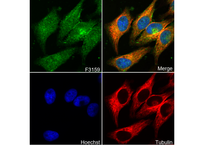 Immunofluorescent analysis of MDA-MB-231 cells using F3159 (green, 1:200), Hoechst (blue) and tubulin (Red).
WB
Validiert von Selleck
-
 Lane 1: Human tonsil, Lane 2: Jurkat, Lane 3: U937, Lane 4: MDA-MB-231