Technische Daten
| Formel | C23H22ClN5O |
|||
| Molekulargewicht | 419.91 | CAS-Nr. | 405168-58-3 | |
| Löslichkeit (25°C)* | In vitro | DMSO | Insoluble | |
| Water | Insoluble | |||
| Ethanol | Insoluble | |||
|
* <1 mg/ml bedeutet schwer löslich oder unlöslich. * Bitte beachten Sie, dass Selleck die Löslichkeit aller Verbindungen intern testet und die tatsächliche Löslichkeit geringfügig von veröffentlichten Werten abweichen kann. Dies ist normal und ist auf geringfügige Batch-zu-Batch-Variationen zurückzuführen. * Versand bei Raumtemperatur (Stabilitätstests zeigen, dass dieses Produkt ohne Kühlmaßnahmen versendet werden kann.) |
||||
Vorbereitung von Stammlösungen
Biologische Aktivität
| Beschreibung | CHIR-124 ist ein neuartiger und potenter Chk1-Inhibitor mit einer IC50 von 0,3 nM in einem zellfreien Assay. Er zeigt eine 2.000-fache Selektivität gegenüber Chk2 und eine 500- bis 5.000-fach geringere Aktivität gegenüber CDK2/4 und Cdc2. | ||||||||
|---|---|---|---|---|---|---|---|---|---|
| Ziele |
|
||||||||
| In vitro | CHIR-124 ist ein chinolonbasiertes kleines Molekül, das strukturell nicht mit anderen bekannten Inhibitoren von Chk1 verwandt ist. Diese Verbindung interagiert synergistisch mit Topoisomerase-Giften (z.B. Camptothecin oder SN-38) und führt zu einer Wachstumshemmung in einer Vielzahl von Krebszelllinien, einschließlich Brustkarzinom (MDA-MB-231 und MDA-MB-435) und Darmkarzinom (SW-620 und Colo205), die alle das mutierte p53-Gen enthalten. Es hebt die SN-38-induzierten S- und G2-M Cell Cycle Checkpoints auf und verstärkt die Apoptose in MDA-MD-435 Brustkrebszellen. Die Aufhebung des G2-M Cell Cycle Checkpoints und die Induktion der Apoptose durch diese Chemikalie werden durch den Verlust von p53 verstärkt. Diese Verbindung zielt auch potent auf andere Kinasen wie PDGFR und Flt3 mit IC50 von 6,6 nM bzw. 5,8 nM ab. |
||||||||
| In vivo | CHIR-124 verstärkt die wachstumshemmenden Effekte, indem es den G2-M Cell Cycle Checkpoint aufhebt und die Tumorapoptose in einem orthotopen Brustkrebs-Xenograftmodell erhöht. |
Protokoll (aus Referenz)
| Kinase-Assay: |
|
|---|---|
| Zell-Assay: |
|
| Tierstudie: |
|
Referenzen
|
Kundenproduktvalidierung
-
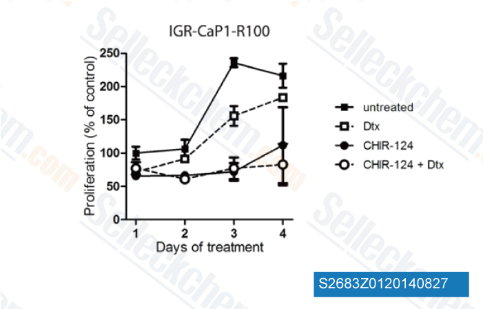
Daten von [ Oncotarget , 2014 , 5(3), 667-78 ]
-
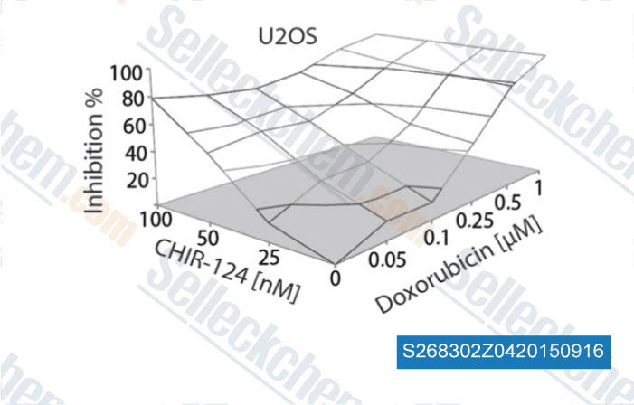
Daten von [ , , J Pathol, 2015, 236: 348-359 ]
-
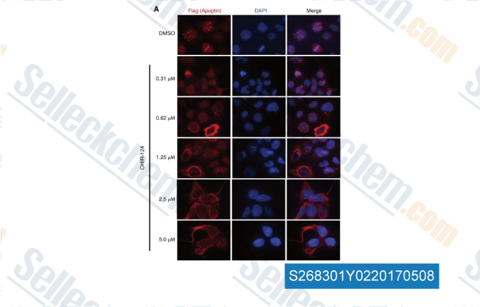
Daten von [ , , J Virol, 2016, 90(20):9433-45 ]
-
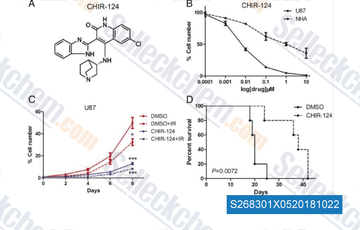
Daten von [ , , Transl Oncol, 2018, 11(1):140-146 ]
Sellecks CHIR-124 Wurde zitiert von 47 Publikationen
| A patient-derived T cell lymphoma biorepository uncovers pathogenetic mechanisms and host-related therapeutic vulnerabilities [ Cell Rep Med, 2025, S2666-3791(25)00102-8] | PubMed: 40147445 |
| The mitotic ATR-Chk1 pathway promotes CDK1 activity for faithful chromosome segregation [ Cell Rep, 2025, 44(8):116019] | PubMed: 40705605 |
| Enhancing transcription-replication conflict targets ecDNA-positive cancers [ Nature, 2024, 635(8037):210-218] | PubMed: 39506153 |
| The MYCN oncoprotein is an RNA-binding accessory factor of the nuclear exosome targeting complex [ Mol Cell, 2024, S1097-2765(24)00285-5] | PubMed: 38703770 |
| Replicative senescence is ATM driven, reversible, and accelerated by hyperactivation of ATM at normoxia [ bioRxiv, 2024, 2024.06.24.600514] | PubMed: 38979390 |
| p53-independent tumor suppression by cell-cycle arrest via CREB/ATF transcription factor OASIS [ Cell Rep, 2023, S2211-1247(23)00490-4] | PubMed: 37178686 |
| The MRN complex maintains the biliary-derived hepatocytes in liver regeneration through ATR-Chk1 pathway [ NPJ Regen Med, 2023, 8(1):20] | PubMed: 37024481 |
| The MRN complex maintains the biliary-derived hepatocytes in liver regeneration through ATR-Chk1 pathway [ npj Regenerative Medicine, 2023, 20-2023)] | PubMed: None |
| Alternative Lengthening of Telomeres in Pediatric High-Grade Glioma and Therapeutic Implications [ Cancers (Basel), 2023, 15(12)3070] | PubMed: 37370681 |
| The Autonomous Parvovirus Minute Virus of Mice Localizes to Cellular Sites of DNA Damage Using ATR Signaling [ Viruses, 2023, 15(6)1243] | PubMed: 37376543 |
RÜCKGABERICHTLINIE
Die bedingungslose Rückgaberichtlinie von Selleck Chemical gewährleistet unseren Kunden ein reibungsloses Online-Einkaufserlebnis. Wenn Sie in irgendeiner Weise mit Ihrem Kauf unzufrieden sind, können Sie jeden Artikel innerhalb von 7 Tagen nach Erhalt zurückgeben. Im Falle von Produktqualitätsproblemen, sei es protokollbezogene oder produktbezogene Probleme, können Sie jeden Artikel innerhalb von 365 Tagen ab dem ursprünglichen Kaufdatum zurückgeben. Bitte befolgen Sie die nachstehenden Anweisungen, wenn Sie Produkte zurücksenden.
VERSAND UND LAGERUNG
Selleck-Produkte werden bei Raumtemperatur transportiert. Wenn Sie das Produkt bei Raumtemperatur erhalten, seien Sie versichert, dass die Qualitätskontrollabteilung von Selleck Experimente durchgeführt hat, um zu überprüfen, dass die normale Temperaturplatzierung von einem Monat die biologische Aktivität von Pulverprodukten nicht beeinträchtigt. Nach dem Sammeln lagern Sie das Produkt bitte gemäß den in der Datenblatt beschriebenen Anforderungen. Die meisten Selleck-Produkte sind unter den empfohlenen Bedingungen stabil.
NICHT FÜR DIE ANWENDUNG AM MENSCHEN, FÜR VETERINÄRMEDIZINISCHE DIAGNOSTIK ODER THERAPEUTISCHE ZWECKE.