|
Wie zu zitieren 1. Für Zitate im Text (Materialien & Methoden): 2. Für die Tabelle der Schlüsselressourcen: |
||
|
Gebührenfrei: (877) 796-6397 -- Nur USA und Kanada -- |
Fax: +1-832-582-8590 Bestellungen: +1-832-582-8158 |
Technischer Support: +1-832-582-8158 Ext:3 Bitte geben Sie Ihre Bestellnummer in der E-Mail an. Wir bemühen uns, alle E-Mail-Anfragen innerhalb eines Werktages zu beantworten. |
Biologische Beschreibung
| Spezifität | Collagen IV Antibody [E5G18] weist endogene Spiegel des gesamten Collagen IV-Proteins nach. |
|---|---|
| Hintergrund | Collagen IV ist das primäre strukturelle Kollagen von Basalmembranen, spezialisierten extrazellulären Matrizen, die mechanische Unterstützung bieten, die Zelladhäsion regulieren und andere Matrixkomponenten organisieren. Es ist ein Heterotrimer, das aus Kombinationen von sechs α-Ketten (α1–α6) besteht, die von verschiedenen Genen kodiert werden und typischerweise in den meisten Geweben als zwei α1- und eine α2-Kette angeordnet sind, die intrazellulär zu tripelhelikalen Protomeren assemblieren. Diese Protomeren werden sezerniert und über ihre NC1- und 7S-Domänen vernetzt, um ein stabiles, blattartiges Netzwerk zu bilden, das Laminine, Proteoglykane und Wachstumsfaktoren verankert. Collagen IV wird in epithelialen und endothelialen Basalmembranen mit gewebespezifischer Isoformzusammensetzung weit verbreitet exprimiert und spielt eine wesentliche Rolle bei Entwicklung, Gewebeintegrität, Zellmigration und Signalübertragung durch Integrin wie α1β1 und α2β1. Defekte oder abnormale Vernetzung von Collagen IV tragen zu verschiedenen Pathologien bei, darunter Alport-Syndrom, Goodpasture-Syndrom, diabetische Nephropathie und bestimmte dermatologische Erkrankungen. |
Nutzungsinformationen
| Anwendung | IHC | Verdünnung |
|
||
|---|---|---|---|---|---|
| Reaktivität | Human | ||||
| Quelle | Mouse Monoclonal Antibody | MW | |||
| Lagerpuffer | PBS, pH 7.2+50% Glycerol+0.05% BSA+0.01% NaN3 | Lagerung (Ab dem Datum des Erhalts) |
-20°C (avoid freeze-thaw cycles), 2 years | ||
| IHC |
Experimental Protocol:
Deparaffinization/Rehydration
1. Deparaffinize/hydrate sections:
2. Incubate sections in three washes of xylene for 5 min each.
3. Incubate sections in two washes of 100% ethanol for 10 min each.
4. Incubate sections in two washes of 95% ethanol for 10 min each.
5. Wash sections two times in dH2O for 5 min each.
6.Antigen retrieval: For Citrate: Heat slides in a microwave submersed in 1X citrate unmasking solution until boiling is initiated; continue with 10 min at a sub-boiling temperature (95°-98°C). Cool slides on bench top for 30 min.
Staining
1. Wash sections in dH2O three times for 5 min each.
2. Incubate sections in 3% hydrogen peroxide for 10 min.
3. Wash sections in dH2O two times for 5 min each.
4. Wash sections in wash buffer for 5 min.
5. Block each section with 100–400 µl of blocking solution for 1 hr at room temperature.
6. Remove blocking solution and add 100–400 µl primary antibody diluent in to each section. Incubate overnight at 4°C.
7. Remove antibody solution and wash sections with wash buffer three times for 5 min each.
8. Cover section with 1–3 drops HRPas needed. Incubate in a humidified chamber for 30 min at room temperature.
9. Wash sections three times with wash buffer for 5 min each.
10. Add DAB Chromogen Concentrate to DAB Diluent and mix well before use.
11. Apply 100–400 µl DAB to each section and monitor closely. 1–10 min generally provides an acceptable staining intensity.
12. Immerse slides in dH2O.
13. If desired, counterstain sections with hematoxylin.
14. Wash sections in dH2O two times for 5 min each.
15. Dehydrate sections: Incubate sections in 95% ethanol two times for 10 sec each; Repeat in 100% ethanol, incubating sections two times for 10 sec each; Repeat in xylene, incubating sections two times for 10 sec each.
16. Mount sections with coverslips and mounting medium.
|
Referenzen
|
Anwendungsdaten
IHC
Validiert von Selleck
-
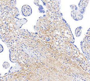 Immunohistochemical analysis of formalin fixed paraffin embedded human placenta tissue with F1667 at 1:125 dilution.