|
Wie zu zitieren 1. Für Zitate im Text (Materialien & Methoden): 2. Für die Tabelle der Schlüsselressourcen: |
||
|
Gebührenfrei: (877) 796-6397 -- Nur USA und Kanada -- |
Fax: +1-832-582-8590 Bestellungen: +1-832-582-8158 |
Technischer Support: +1-832-582-8158 Ext:3 Bitte geben Sie Ihre Bestellnummer in der E-Mail an. Wir bemühen uns, alle E-Mail-Anfragen innerhalb eines Werktages zu beantworten. |
Biologische Beschreibung
| Spezifität | Glutathione Antibody [A11G7] detektiert endogene Spiegel des gesamten Glutathione-Proteins. |
|---|---|
| Hintergrund | Glutathione (GSH) ist ein niedermolekulares Tripeptid-Thiol-Antioxidans, das aus Glutamat, Cystein und Glycin besteht und über zwei aufeinanderfolgende ATP-abhängige Schritte synthetisiert wird, die durch Glutamat-Cystein-Ligase und Glutathion-Synthetase katalysiert werden. Es existiert primär in reduzierter (GSH) und oxidierter Disulfid (GSSG)-Form, die sich gegenseitig umwandeln, um das zelluläre Redoxgleichgewicht zu regulieren. GSH weist eine einzigartige γ-Glutamyl-Peptidbindung zwischen der γ-Carboxylgruppe von Glutamat und der Aminogruppe von Cystein auf, die eine Resistenz gegen den Abbau durch γ-Glutamyl-Cyclotransferase und die meisten Peptidasen verleiht; das Cystein-Thiol (-SH) fungiert als nukleophiles Zentrum für die Redoxchemie, während Glycin den C-Terminus stabilisiert. GSH fängt reaktive Sauerstoff- und Stickstoffspezies (ROS/RNS) direkt über Thiol-Disulfid-Austausch ab, dient als essentielles Reduktionsmittel für Glutathion-Peroxidasen (GPx), die Peroxide entgiften (wobei GSSG durch Glutathion-Reduktase unter Verwendung von NADPH wieder zu GSH reduziert wird), und konjugiert elektrophile Xenobiotika, Schwermetalle und endogene Toxine über Glutathion-S-Transferasen (GSTs) zur Phase-II-Entgiftung und zum Mercaptursäure-Export. Diese Prozesse erhalten die Sulfhydryl-Homöostase von Proteinen, unterstützen die Nrf2-gesteuerte antioxidative Genexpression, erleichtern die Eisen-Schwefel-Cluster-Montage und den Metalltransport und modulieren Signalwege wie NF-κB. GSH-Depletion ist an oxidativem Stress-bedingten Krankheiten beteiligt, einschließlich Parkinson-Krankheit, Leberzirrhose, Krebs, Diabetes und vorzeitiger Alterung. |
Nutzungsinformationen
| Anwendung | IF, FCM | Verdünnung |
|
||||
|---|---|---|---|---|---|---|---|
| Reaktivität | |||||||
| Quelle | Mouse Monoclonal Antibody | MW | |||||
| Lagerpuffer | PBS, pH 7.2+50% Glycerol+0.05% BSA+0.01% NaN3 | Lagerung (Ab dem Datum des Erhalts) |
-20°C (avoid freeze-thaw cycles), 2 years | ||||
| IF |
Experimental Protocol:
Sample Preparation
1. Adherent Cells: Place a clean, sterile coverslip in a culture dish. Once the cells grow to near confluence as a monolayer, remove the coverslip for further use.
2. Suspension Cells: Seed the cells onto a clean, sterile slide coated with poly-L-lysine.
3. Frozen Sections: Allow the slide to thaw at room temperature. Wash it with pure water or PBS for 2 times, 3 minutes each time.
4. Paraffin Sections: Deparaffinization and rehydration. Wash the slide with pure water or PBS for 3 times, 3 minutes each time. Then perform antigen retrieval.
Fixation
1. Fix the cell coverslips/spots or tissue sections at room temperature using a fixative such as 4% paraformaldehyde (4% PFA) for 10-15 minutes.
2. Wash the sample with PBS for 3 times, 3 minutes each time.
Permeabilization
1.Add a detergent such as 0.1–0.3% Triton X-100 to the sample and incubate at room temperature for 10–20 minutes.
(Note: This step is only required for intracellular antigens. For antigens expressed on the cell membrane, this step is unnecessary.)
Wash the sample with PBS for 3 times, 3 minutes each time.
Blocking
Add blocking solution and incubate at room temperature for at least 1 hour. (Common blocking solutions include: serum from the same source as the secondary antibody, BSA, or goat serum.)
Note: Ensure the sample remains moist during and after the blocking step to prevent drying, which can lead to high background.
Immunofluorescence Staining (Day 1)
1. Remove the blocking solution and add the diluted primary antibody.
2. Incubate the sample in a humidified chamber at 4°C overnight.
Immunofluorescence Staining (Day 2)
1. Remove the primary antibody and wash with PBST for 3 times, 5 minutes each time.
2. Add the diluted fluorescent secondary antibody and incubate in the dark at 4°C for 1–2 hours.
3. Remove the secondary antibody and wash with PBST for 3 times, 5 minutes each time.
4. Add diluted DAPI and incubate at room temperature in the dark for 5–10 minutes.
5. Wash with PBST for 3 times, 5 minutes each time.
Mounting
1. Mount the sample with an anti-fade mounting medium.
2. Allow the slide to dry at room temperature overnight in the dark.
3. Store the slide in a slide storage box at 4°C, protected from light.
|
Referenzen
|
Anwendungsdaten
IF
Validiert von Selleck
-
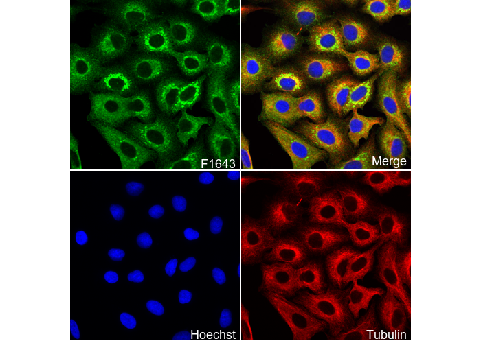 Immunofluorescent analysis of A549 cells using F1643 (green, 1:100), Hoechst (blue) and tubulin (Red).