|
Wie zu zitieren 1. Für Zitate im Text (Materialien & Methoden): 2. Für die Tabelle der Schlüsselressourcen: |
||
|
Gebührenfrei: (877) 796-6397 -- Nur USA und Kanada -- |
Fax: +1-832-582-8590 Bestellungen: +1-832-582-8158 |
Technischer Support: +1-832-582-8158 Ext:3 Bitte geben Sie Ihre Bestellnummer in der E-Mail an. Wir bemühen uns, alle E-Mail-Anfragen innerhalb eines Werktages zu beantworten. |
Biologische Beschreibung
| Spezifität | Glutathione Peroxidase 1 Antibody [M21J11] detektiert endogene Spiegel des gesamten Glutathione Peroxidase 1 Proteins. |
|---|---|
| Hintergrund | Glutathione Peroxidase 1 (GPx1) ist das wichtigste intrazelluläre Selenoprotein-Antioxidans-Enzym, das Zellen vor oxidativen Schäden schützt, indem es Wasserstoffperoxid und Lipidhydroperoxide zu Wasser und Alkoholen reduziert, wobei reduziertes Glutathione (GSH) als Elektronendonor verwendet wird; das resultierende oxidierte Glutathiondisulfid (GSSG) wird durch Glutathionreduktase recycelt. GPx1 ist ein Homotetramer aus vier identischen 22–23 kDa Untereinheiten, die jeweils vom GPX1-Gen (Chromosom 3p21.31) kodiert werden und Selenocystein (Sec) am aktiven Zentrum durch UGA-Codon-Rekodierung, gesteuert durch das SECIS-Element in der mRNA 3′ UTR, einbauen. Die katalytische Tetrade (Sec-Gln-Trp-Asn) befindet sich in einer Tasche, die von konservierten Arginin- und Lysinresten benachbarter Untereinheiten gebildet wird, was die GSH-Bindung erleichtert und eine Ping-Pong-Bisubstratkinetik für eine effiziente Peroxid-Entgiftung ermöglicht. Sowohl zytosolische als auch mitochondriale GPx1 erhalten die Redox-Homöostase aufrecht, indem sie mit Superoxiddismutase (SOD) und Katalase zusammenarbeiten, um reaktive Sauerstoffspezies (ROS) zu neutralisieren und so Lipidperoxidation, Proteincarbonylierung, DNA-Schäden und aberrante H2O2-vermittelte Signalgebung zu verhindern, die Apoptose, Wachstumsfaktorreaktionen, Insulinsensitivität und Nrf2-abhängige Genexpression beeinflussen können. GPx1-Mangel ist mit Herz-Kreislauf-Erkrankungen (Atherosklerose, Endothelfunktionsstörung), Neurodegeneration (wie durch Neuroprotektion bei Schlaganfall/traumatischer Hirnverletzung gezeigt) und Krebs (wo seine Rolle entweder tumorfördernd oder risikomodulierend sein kann, beeinflusst durch den Pro198Leu-Polymorphismus) verbunden. GPx1 dient auch als wichtiger Biomarker für den Selenstatus und wird transkriptionell durch Nrf2, NF-κB und p53 reguliert. |
Nutzungsinformationen
| Anwendung | WB, IHP, FCM | Verdünnung |
|
||||||
|---|---|---|---|---|---|---|---|---|---|
| Reaktivität | Human | ||||||||
| Quelle | Rabbit Monoclonal Antibody | MW | 22 kDa | ||||||
| Lagerpuffer | PBS, pH 7.2+50% Glycerol+0.05% BSA+0.01% NaN3 | Lagerung (Ab dem Datum des Erhalts) |
-20°C (avoid freeze-thaw cycles), 2 years | ||||||
| WB |
Experimental Protocol:
Sample preparation
1. Tissue: Lyse the tissue sample by adding an appropriate volume of ice-cold RIPA/NP-40 Lysis Buffer (containing Protease Inhibitor Cocktail),and homogenize the tissue at a low temperature. 2. Adherent cell: Aspirate the culture medium and wash the cells with ice-cold PBS twice. Lyse the cells by adding an appropriate volume of RIPA/NP-40 Lysis Buffer (containing Protease Inhibitor Cocktail) and put the sample on ice for 5 min. 3. Suspension cell: Transfer the culture medium to a pre-cooled centrifuge tube. Centrifuge and aspirate the supernatant. Wash the cells with ice-cold PBS twice. Lyse the cells by adding an appropriate volume of RIPA/NP-40 Lysis Buffer (containing Protease Inhibitor Cocktail) and put the sample on ice for 5 min. 4. Place the lysate into a pre-cooled microcentrifuge tube. Centrifuge at 4°C for 15 min. Collect the supernatant;
5. Remove a small volume of lysate to determine the protein concentration;
6. Combine the lysate with protein loading buffer. Boil 20 µL sample under 95-100°C for 5 min. Centrifuge for 5 min after cool down on ice.
Electrophoretic separation
1. According to the concentration of extracted protein, load appropriate amount of protein sample and marker onto SDS-PAGE gels for electrophoresis. Recommended separating gel (lower gel) concentration: 10%. Reference Table for Selecting SDS-PAGE Separation Gel Concentrations 2. Power up 80V for 30 minutes. Then the power supply is adjusted (110 V~150 V), the Marker is observed, and the electrophoresis can be stopped when the indicator band of the predyed protein Marker where the protein is located is properly separated. (Note that the current should not be too large when electrophoresis, too large current (more than 150 mA) will cause the temperature to rise, affecting the result of running glue. If high currents cannot be avoided, an ice bath can be used to cool the bath.)
Transfer membrane
1. Take out the converter, soak the clip and consumables in the pre-cooled converter;
2. Activate PVDF membrane with methanol for 1 min and rinse with transfer buffer;
3. Install it in the order of "black edge of clip - sponge - filter paper - filter paper - glue -PVDF membrane - filter paper - filter paper - sponge - white edge of clip"; 4. The protein was electrotransferred to PVDF membrane. ( 0.45 µm PVDF membrane is recommended ) Reference Table for Selecting PVDF Membrane Pore Size Specifications Recommended conditions for wet transfer: 200 mA, 60 min. ( Note that the transfer conditions can be adjusted according to the protein size. For high-molecular-weight proteins, a higher current and longer transfer time are recommended. However, ensure that the transfer tank remains at a low temperature to prevent gel melting.)
Block
1. After electrotransfer, wash the film with TBST at room temperature for 5 minutes;
2. Incubate the film in the blocking solution for 1 hour at room temperature;
3. Wash the film with TBST for 3 times, 5 minutes each time.
Antibody incubation
1. Use 5% skim milk powder to prepare the primary antibody working liquid (recommended dilution ratio for primary antibody 1:1000), gently shake and incubate with the film at 4°C overnight; 2. Wash the film with TBST 3 times, 5 minutes each time;
3. Add the secondary antibody to the blocking solution and incubate with the film gently at room temperature for 1 hour;
4. After incubation, wash the film with TBST 3 times for 5 minutes each time.
Antibody staining
1. Add the prepared ECL luminescent substrate (or select other color developing substrate according to the second antibody) and mix evenly;
2. Incubate with the film for 1 minute, remove excess substrate (keep the film moist), wrap with plastic film, and expose in the imaging system.
|
Referenzen
|
Anwendungsdaten
WB
Validiert von Selleck
-
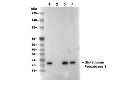 Lane 1: THP1, Lane 2: THP1 (KO GPX1), Lane 3: HL60, Lane 4: Rat liver