|
Wie zu zitieren 1. Für Zitate im Text (Materialien & Methoden): 2. Für die Tabelle der Schlüsselressourcen: |
||
|
Gebührenfrei: (877) 796-6397 -- Nur USA und Kanada -- |
Fax: +1-832-582-8590 Bestellungen: +1-832-582-8158 |
Technischer Support: +1-832-582-8158 Ext:3 Bitte geben Sie Ihre Bestellnummer in der E-Mail an. Wir bemühen uns, alle E-Mail-Anfragen innerhalb eines Werktages zu beantworten. |
Biologische Beschreibung
| Spezifität | HEF1/NEDD9 Antikörper [M16B3] detektiert endogene Spiegel des gesamten HEF1/NEDD9 Proteins. |
|---|---|
| Hintergrund | HEF1, auch bekannt als NEDD9 oder Cas-L, ist ein wichtiges Multidomänen-Adaptorprotein innerhalb der Cas-Familie, das die Integrin-vermittelte Signalgebung an fokalen Adhäsionen während der Interphase koordiniert und in der G2/M-Phase an Zentrosomen und mitotischen Strukturen lokalisiert, um Zelladhäsion, Migration, Polarität und Überleben zu regulieren. HEF1 enthält eine N-terminale SH3-Domäne (Aminosäurereste 3–65) zum Binden von prolinreichen Proteinen, eine Substratdomäne (Aminosäurereste 110–356), die mit Tyrosinmotiven wie Y189 und Y214 angereichert ist, die durch Src und FAK phosphoryliert werden, um SH2-Domänen-enthaltende Effektoren wie Crk und Abl zu rekrutieren, eine serinreiche Region (Aminosäurereste 357–560), die ein Vier-Helix-Bündel für strukturelle Stabilität und Proteininteraktionen bildet, und eine C-terminale Fokale Adhäsions-Targeting (FAT)-Domäne (Aminosäurereste 561–834) mit Helix-Loop-Helix-Motiven, die das Andocken von FAK, die Bindung von Src und die Dimerisierung erleichtern. HEF1 integriert Signale von FAK, Src, PDGFR und Abl, was zur Phosphorylierung seiner Substratdomäne und zur Bildung von Signalkomplexen führt, die die Umgestaltung des Aktinzytoskeletts, den Umsatz fokaler Adhäsionen, die Invadopodienbildung über MMP9-Sekretion und die Aktivierung von Signalwegen wie FAK-Src-Aurora-A für Chemotaxis und mitotische Progression fördern; seine Phosphorylierungsdynamik reguliert ferner die Apoptoseresistenz und die Epithel-Mesenchymale Transition. Die Überexpression von NEDD9 fördert die Metastasierung bei Krebsarten wie Glioblastom, Melanom und Lungenkarzinom, indem sie die Invasion und Überlebenssignalgebung verstärkt, während ein Mangel die Tumorprogression und die Leukozytenmigration beeinträchtigt. |
Nutzungsinformationen
| Anwendung | WB, IP | Verdünnung |
|
||||
|---|---|---|---|---|---|---|---|
| Reaktivität | Human, Mouse, Rat, Monkey | ||||||
| Quelle | Mouse Monoclonal Antibody | MW | 105 kDa, 115 kDa | ||||
| Lagerpuffer | PBS, pH 7.2+50% Glycerol+0.05% BSA+0.01% NaN3 | Lagerung (Ab dem Datum des Erhalts) |
-20°C (avoid freeze-thaw cycles), 2 years | ||||
| WB |
Experimental Protocol:
Sample preparation
1. Tissue: Lyse the tissue sample by adding an appropriate volume of ice-cold RIPA/NP-40 Lysis Buffer (containing Protease Inhibitor Cocktail),and homogenize the tissue at a low temperature. 2. Adherent cell: Aspirate the culture medium and wash the cells with ice-cold PBS twice. Lyse the cells by adding an appropriate volume of RIPA/NP-40 Lysis Buffer (containing Protease Inhibitor Cocktail) and put the sample on ice for 5 min. 3. Suspension cell: Transfer the culture medium to a pre-cooled centrifuge tube. Centrifuge and aspirate the supernatant. Wash the cells with ice-cold PBS twice. Lyse the cells by adding an appropriate volume of RIPA/NP-40 Lysis Buffer (containing Protease Inhibitor Cocktail) and put the sample on ice for 5 min. 4. Place the lysate into a pre-cooled microcentrifuge tube. Centrifuge at 4°C for 15 min. Collect the supernatant;
5. Remove a small volume of lysate to determine the protein concentration;
6. Combine the lysate with protein loading buffer. Boil 20 µL sample under 95-100°C for 5 min. Centrifuge for 5 min after cool down on ice.
Electrophoretic separation
1. According to the concentration of extracted protein, load appropriate amount of protein sample and marker onto SDS-PAGE gels for electrophoresis. Recommended separating gel (lower gel) concentration: 5%. Reference Table for Selecting SDS-PAGE Separation Gel Concentrations 2. Power up 80V for 30 minutes. Then the power supply is adjusted (110 V~150 V), the Marker is observed, and the electrophoresis can be stopped when the indicator band of the predyed protein Marker where the protein is located is properly separated. (Note that the current should not be too large when electrophoresis, too large current (more than 150 mA) will cause the temperature to rise, affecting the result of running glue. If high currents cannot be avoided, an ice bath can be used to cool the bath.)
Transfer membrane
1. Take out the converter, soak the clip and consumables in the pre-cooled converter;
2. Activate PVDF membrane with methanol for 1 min and rinse with transfer buffer;
3. Install it in the order of "black edge of clip - sponge - filter paper - filter paper - glue -PVDF membrane - filter paper - filter paper - sponge - white edge of clip"; 4. The protein was electrotransferred to PVDF membrane. ( 0.45 µm PVDF membrane is recommended ) Reference Table for Selecting PVDF Membrane Pore Size Specifications Recommended conditions for wet transfer: 200 mA, 120 min. ( Note that the transfer conditions can be adjusted according to the protein size. For high-molecular-weight proteins, a higher current and longer transfer time are recommended. However, ensure that the transfer tank remains at a low temperature to prevent gel melting.)
Block
1. After electrotransfer, wash the film with TBST at room temperature for 5 minutes;
2. Incubate the film in the blocking solution for 1 hour at room temperature;
3. Wash the film with TBST for 3 times, 5 minutes each time.
Antibody incubation
1. Use 5% skim milk powder to prepare the primary antibody working liquid (recommended dilution ratio for primary antibody 1:1000), gently shake and incubate with the film at 4°C overnight; 2. Wash the film with TBST 3 times, 5 minutes each time;
3. Add the secondary antibody to the blocking solution and incubate with the film gently at room temperature for 1 hour;
4. After incubation, wash the film with TBST 3 times for 5 minutes each time.
Antibody staining
1. Add the prepared ECL luminescent substrate (or select other color developing substrate according to the second antibody) and mix evenly;
2. Incubate with the film for 1 minute, remove excess substrate (keep the film moist), wrap with plastic film, and expose in the imaging system.
|
Referenzen
|
Anwendungsdaten
WB
Validiert von Selleck
-
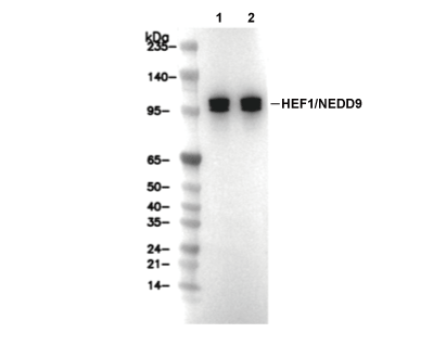 Lane 1: A549, Lane 2: C2C12