|
Wie zu zitieren 1. Für Zitate im Text (Materialien & Methoden): 2. Für die Tabelle der Schlüsselressourcen: |
||
|
Gebührenfrei: (877) 796-6397 -- Nur USA und Kanada -- |
Fax: +1-832-582-8590 Bestellungen: +1-832-582-8158 |
Technischer Support: +1-832-582-8158 Ext:3 Bitte geben Sie Ihre Bestellnummer in der E-Mail an. Wir bemühen uns, alle E-Mail-Anfragen innerhalb eines Werktages zu beantworten. |
Biologische Beschreibung
| Spezifität | Histone H4 (tri methyl Lys20) Antibody [J13H2] erkennt endogene Histon-H4-Proteine nur, wenn diese an Lys20 trimethyliert sind. Dieser Antikörper kreuzreagiert nicht mit nicht-methyliertem, mono-methyliertem oder di-methyliertem Histon H4 Lys20. Dieser Antikörper detektiert ein 95 kDa großes, unspezifisches Protein unbekannten Ursprungs. |
|---|---|
| Hintergrund | Tri-Methyl-Histone H4 (Lys20), auch bekannt als H4K20me3, ist eine spezifische posttranslationale Histonmodifikation, die entscheidend für die Aufrechterhaltung der genomischen Integrität und die Regulierung der Chromatin-Struktur ist. Die Histon-H4-Lysin-20-Methylierung existiert in drei Zuständen: Mono-Methylierung (H4K20me1), Di-Methylierung (H4K20me2) und Tri-Methylierung (H4K20me3), wobei H4K20me3 die Trimethylierungsmarkierung am Lysin-20-Rest von Histon H4 ist. Diese Modifikation ist von Hefe bis zum Menschen evolutionär konserviert und dient als Marker für transkriptionell inaktives Heterochromatin, was auf Regionen des Genoms hinweist, die nicht aktiv transkribiert werden. H4K20me3 ist an spezifischen genomischen Orten wie perizentrischem Heterochromatin, Telomeren, geprägten Regionen und repetitiven Elementen stark angereichert und spielt eine Rolle bei der Aufrechterhaltung dieser Bereiche in einem reprimierten Zustand. Es ist sowohl in Abwesenheit als auch in Anwesenheit von genotoxischem Stress für die genomische Integrität unerlässlich und bildet einen Teil des umfassenderen DNA-Schadensreaktionsnetzwerks (DDR). Die Enzyme SUV4-20H1 und SUV4-20H2 vermitteln hauptsächlich die Tri-Methylierung von H4K20. Die verschiedenen Methylierungszustände von H4K20 ändern sich dynamisch während des Zellzyklus. H4K20me3 bleibt stabiler und zeigt weniger dramatische Änderungen im Vergleich zu H4K20me1. H4K20me3 verstärkt die Chromatinfaltung und spielt eine strukturelle Rolle im Chromatin-Gerüst. Die richtige Regulierung der H4K20-Methylierung ist entscheidend für die Aufrechterhaltung der Chromatin-Struktur und -Funktion, da Störungen dieser Regulierung zu genomischer Instabilität und anderen zellulären Dysfunktionen führen. |
Nutzungsinformationen
| Anwendung | WB, ChIP | Verdünnung |
|
||||||
|---|---|---|---|---|---|---|---|---|---|
| Reaktivität | Human, Mouse, Rat, Monkey, Xenopus, Bovine, Pig | ||||||||
| Quelle | Rabbit Monoclonal Antibody | MW | 11 Kda | ||||||
| Lagerpuffer | PBS, pH 7.2+50% Glycerol+0.05% BSA+0.01% NaN₃ | Lagerung (Ab dem Datum des Erhalts) |
–20°C (avoid freeze-thaw cycles), 2 years | ||||||
| WB |
Experimental Protocol:
Sample preparation
1. Tissue: Lyse the tissue sample by adding an appropriate volume of ice-cold RIPA/Nuclear Lysis Buffer (containing Protease Inhibitor Cocktail),and homogenize the tissue at a low temperature or lyse it by sonication on ice, then incubate on ice for 30 minutes. 2. Adherent cell: Aspirate the culture medium and transfer the cells into an EP tube. Wash the cells with ice-cold PBS twice. Add an appropriate volume of RIPA/Nuclear Lysis Buffer (containing Protease Inhibitor Cocktail), sonicate to lyse the cells, and incubate on ice for 30 minutes. 3. Suspension cell: Transfer the culture medium to a pre-cooled centrifuge tube. Centrifuge and aspirate the supernatant. Wash the cells with ice-cold PBS twice.Add an appropriate volume of RIPA/Nuclear Lysis Buffer (containing Protease Inhibitor Cocktail), sonicate to lyse the cells, and incubate on ice for 30 minutes. 4. Place the lysate into a pre-cooled microcentrifuge tube. Centrifuge at 4°C for 15 min. Collect the supernatant;
5. Remove a small volume of lysate to determine the protein concentration;
6. Combine the lysate with protein loading buffer. Boil 20 µL sample under 95-100°C for 5 min. Centrifuge for 5 min after cool down on ice.
Electrophoretic separation
1. According to the concentration of extracted protein, load appropriate amount of protein sample and marker onto SDS-PAGE gels for electrophoresis. Recommended separating gel (lower gel) concentration: 20%. Reference Table for Selecting SDS-PAGE Separation Gel Concentrations 2. Power up 80V for 30 minutes. Then the power supply is adjusted (110 V~150 V), the Marker is observed, and the electrophoresis can be stopped when the indicator band of the predyed protein Marker where the protein is located is properly separated. (Note that the current should not be too large when electrophoresis, too large current (more than 150 mA) will cause the temperature to rise, affecting the result of running glue. If high currents cannot be avoided, an ice bath can be used to cool the bath.)
Transfer membrane
1. Take out the converter, soak the clip and consumables in the pre-cooled converter;
2. Activate PVDF membrane with methanol for 1 min and rinse with transfer buffer;
3. Install it in the order of "black edge of clip - sponge - filter paper - filter paper - glue -PVDF membrane - filter paper - filter paper - sponge - white edge of clip"; 4. The protein was electrotransferred to PVDF membrane. ( 0.22 µm PVDF membrane is recommended )) Reference Table for Selecting PVDF Membrane Pore Size Specifications Recommended conditions for wet transfer: 200 mA, 60 min. ( Note that the transfer conditions can be adjusted according to the protein size. For high-molecular-weight proteins, a higher current and longer transfer time are recommended. However, ensure that the transfer tank remains at a low temperature to prevent gel melting.)
Block
1. After electrotransfer, wash the film with TBST at room temperature for 5 minutes;
2. Incubate the film in the blocking solution for 1 hour at room temperature;
3. Wash the film with TBST for 3 times, 5 minutes each time.
Antibody incubation
1. Use 5% skim milk powder to prepare the primary antibody working liquid (recommended dilution ratio for primary antibody 1:1000), gently shake and incubate with the film at 4°C overnight; 2. Wash the film with TBST 3 times, 5 minutes each time;
3. Add the secondary antibody to the blocking solution and incubate with the film gently at room temperature for 1 hour;
4. After incubation, wash the film with TBST 3 times for 5 minutes each time.
Antibody staining
575. Add the prepared ECL luminescent substrate (or select other color developing substrate according to the second antibody) and mix evenly;
2. Incubate with the film for 1 minute, remove excess substrate (keep the film moist), wrap with plastic film, and expose in the imaging system.
|
Referenzen
|
Anwendungsdaten
WB
Validiert von Selleck
-
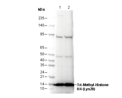 Lane 1: Hela
Lane 2: NIH3T3