|
Wie zu zitieren 1. Für Zitate im Text (Materialien & Methoden): 2. Für die Tabelle der Schlüsselressourcen: |
||
|
Gebührenfrei: (877) 796-6397 -- Nur USA und Kanada -- |
Fax: +1-832-582-8590 Bestellungen: +1-832-582-8158 |
Technischer Support: +1-832-582-8158 Ext:3 Bitte geben Sie Ihre Bestellnummer in der E-Mail an. Wir bemühen uns, alle E-Mail-Anfragen innerhalb eines Werktages zu beantworten. |
Biologische Beschreibung
| Spezifität | JAK3 Antibody [F8D8] weist endogene Spiegel des gesamten JAK3-Proteins nach. |
|---|---|
| Hintergrund | JAK3 (Januskinase 3) ist ein Mitglied der Januskinase (JAK)-Familie der Nicht-Rezeptor-Tyrosinkinasen, bestehend aus JAK1, JAK2, JAK3 und TYK2, und wird überwiegend in hämatopoetischen Zellen exprimiert, wo es spezifisch mit der gemeinsamen Gammakette (γc) der Zytokinrezeptoren für IL-2, IL-4, IL-7, IL-9, IL-15 und IL-21 assoziiert ist. JAK3 weist eine konservierte Multidomänenorganisation auf, die eine N-terminale FERM-Domäne für die Rezeptorbindung, eine SH2-ähnliche Domäne, eine Pseudokinase-Domäne (JH2), der eine intrinsische katalytische Aktivität fehlt, die aber die benachbarte aktive Kinase-Domäne (JH1) negativ reguliert, und eine C-terminale JH1-Kinase-Domäne mit einer zweilappigen Struktur, gekennzeichnet durch eine β-Faltblatt-N-Lobe, eine αC-Helix und eine C-Lobe, die eine eingefügte αFG-Helix (Reste 1029–1038) enthält, aufweist; regulatorische Phosphorylierungsstellen umfassen Tyr980 und Tyr981 in der Aktivierungsschleife von JH1. Zytokin-induzierte Rezeptordimerisierung löst JAK3-Autophosphorylierung und Transphosphorylierung mit JAK1 oder JAK2 aus, wodurch die JH1-Domäne aktiviert wird, um Rezeptortyrosine zu phosphorylieren, die als Andockstellen für STAT-Proteine (wie STAT5) über SH2-Domänen dienen. Nachfolgende STAT-Aktivierung fördert die Gen-Transkription, die für die Proliferation, Differenzierung, das Überleben und die Entwicklung von Immunzellen, insbesondere in T-Zellen, B-Zellen und NK-Zellen, entscheidend ist. Dieser JAK-STAT-Signalweg ist essentiell für die Lymphopoese und adaptive Immunität, wobei die JH2-Pseudokinase-Domäne die JH1-Aktivität bis zur Ligandenbindung allosterisch unterdrückt. Eine Dysregulation von JAK3, einschließlich Gain-of-Function-Mutationen (oft in JH2), führt zu einer anhaltenden Signalgebung und Onkogenese bei Leukämien und Lymphomen, während Loss-of-Function-Mutationen zu schwerer kombinierter Immundefizienz (SCID) führen. |
Nutzungsinformationen
| Anwendung | WB, IP | Verdünnung |
|
||||
|---|---|---|---|---|---|---|---|
| Reaktivität | Human | ||||||
| Quelle | Rabbit Monoclonal Antibody | MW | 115 kDa | ||||
| Lagerpuffer | PBS, pH 7.2+50% Glycerol+0.05% BSA+0.01% NaN3 | Lagerung (Ab dem Datum des Erhalts) |
-20°C (avoid freeze-thaw cycles), 2 years | ||||
| WB |
Experimental Protocol:
Sample preparation
1. Tissue: Lyse the tissue sample by adding an appropriate volume of ice-cold RIPA/NP-40 Lysis Buffer (containing Protease Inhibitor Cocktail),and homogenize the tissue at a low temperature. 2. Adherent cell: Aspirate the culture medium and wash the cells with ice-cold PBS twice. Lyse the cells by adding an appropriate volume of RIPA/NP-40 Lysis Buffer (containing Protease Inhibitor Cocktail) and put the sample on ice for 5 min. 3. Suspension cell: Transfer the culture medium to a pre-cooled centrifuge tube. Centrifuge and aspirate the supernatant. Wash the cells with ice-cold PBS twice. Lyse the cells by adding an appropriate volume of RIPA/NP-40 Lysis Buffer (containing Protease Inhibitor Cocktail) and put the sample on ice for 5 min. 4. Place the lysate into a pre-cooled microcentrifuge tube. Centrifuge at 4°C for 15 min. Collect the supernatant;
5. Remove a small volume of lysate to determine the protein concentration;
6. Combine the lysate with protein loading buffer. Boil 20 µL sample under 95-100°C for 5 min. Centrifuge for 5 min after cool down on ice.
Electrophoretic separation
1. According to the concentration of extracted protein, load appropriate amount of protein sample and marker onto SDS-PAGE gels for electrophoresis. Recommended separating gel (lower gel) concentration: 5%. Reference Table for Selecting SDS-PAGE Separation Gel Concentrations 2. Power up 80V for 30 minutes. Then the power supply is adjusted (110 V~150 V), the Marker is observed, and the electrophoresis can be stopped when the indicator band of the predyed protein Marker where the protein is located is properly separated. (Note that the current should not be too large when electrophoresis, too large current (more than 150 mA) will cause the temperature to rise, affecting the result of running glue. If high currents cannot be avoided, an ice bath can be used to cool the bath.)
Transfer membrane
1. Take out the converter, soak the clip and consumables in the pre-cooled converter;
2. Activate PVDF membrane with methanol for 1 min and rinse with transfer buffer;
3. Install it in the order of "black edge of clip - sponge - filter paper - filter paper - glue -PVDF membrane - filter paper - filter paper - sponge - white edge of clip"; 4. The protein was electrotransferred to PVDF membrane. ( 0.45 µm PVDF membrane is recommended ) Reference Table for Selecting PVDF Membrane Pore Size Specifications Recommended conditions for wet transfer: 200 mA, 120 min. ( Note that the transfer conditions can be adjusted according to the protein size. For high-molecular-weight proteins, a higher current and longer transfer time are recommended. However, ensure that the transfer tank remains at a low temperature to prevent gel melting.)
Block
1. After electrotransfer, wash the film with TBST at room temperature for 5 minutes;
2. Incubate the film in the blocking solution for 1 hour at room temperature;
3. Wash the film with TBST for 3 times, 5 minutes each time.
Antibody incubation
1. Use 5% skim milk powder to prepare the primary antibody working liquid (recommended dilution ratio for primary antibody 1:1000), gently shake and incubate with the film at 4°C overnight; 2. Wash the film with TBST 3 times, 5 minutes each time;
3. Add the secondary antibody to the blocking solution and incubate with the film gently at room temperature for 1 hour;
4. After incubation, wash the film with TBST 3 times for 5 minutes each time.
Antibody staining
1. Add the prepared ECL luminescent substrate (or select other color developing substrate according to the second antibody) and mix evenly;
2. Incubate with the film for 1 minute, remove excess substrate (keep the film moist), wrap with plastic film, and expose in the imaging system.
|
Referenzen
|
Anwendungsdaten
WB
Validiert von Selleck
-
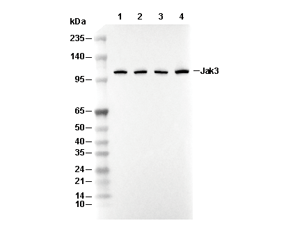 Lane 1: NK-92, Lane 2: Jurkat, Lane 3: SET-2, Lane 4: KARPAS-299