|
Wie zu zitieren 1. Für Zitate im Text (Materialien & Methoden): 2. Für die Tabelle der Schlüsselressourcen: |
||
|
Gebührenfrei: (877) 796-6397 -- Nur USA und Kanada -- |
Fax: +1-832-582-8590 Bestellungen: +1-832-582-8158 |
Technischer Support: +1-832-582-8158 Ext:3 Bitte geben Sie Ihre Bestellnummer in der E-Mail an. Wir bemühen uns, alle E-Mail-Anfragen innerhalb eines Werktages zu beantworten. |
Biologische Beschreibung
| Spezifität | MCP1 Antibody [M3B19] weist endogene Konzentrationen des gesamten MCP1-Proteins nach. |
|---|---|
| Hintergrund | Monozyten-Chemoattraktans-Protein-1 (MCP-1/CCL2) ist ein Mitglied der C-C-Chemokin-Familie und fungiert als potenter chemotaktischer Faktor für Monozyten. Es wird als identisch mit JE angesehen, einem Gen, das ursprünglich in Mausfibroblasten als durch den Plättchen-Wachstumsfaktor induziert identifiziert wurde. Das menschliche MCP-1-Gen befindet sich auf Chromosom 17q11.2 und kodiert ein Protein aus 76 Aminosäuren mit einem Molekulargewicht von etwa 13 kDa. MCP-1 gehört zu einer Unterfamilie von Chemokinen, die mindestens vier Mitglieder umfasst: MCP-1, MCP-2, MCP-3 und MCP-4. CCL2 wird von einer Vielzahl von Zelltypen produziert, entweder konstitutiv oder als Reaktion auf Stimuli wie oxidativen Stress, Zytokine und Wachstumsfaktoren. Seine Quellen umfassen Endothelzellen, Fibroblasten, Epithelzellen, glatte Muskelzellen, Mesangialzellen, Astrozyten, Monozyten und Mikroglia – Zelltypen, die wichtige Rollen bei der antiviralen Immunabwehr sowohl im Kreislauf als auch in Geweben spielen. Funktional steuert CCL2 die Migration und Infiltration von Monozyten, Gedächtnis-T-Zellen und natürlichen Killer (NK)-Zellen. Die biologischen Wirkungen von CCL2 werden über seinen Rezeptor, CCR2, vermittelt, dessen Expression im Vergleich zu der von CCL2 stärker eingeschränkt ist. CCR2 existiert in zwei alternativ gespleißten Isoformen, CCR2A und CCR2B, die sich nur in ihren C-terminalen Regionen unterscheiden. |
Nutzungsinformationen
| Anwendung | IHC | Verdünnung |
|
|---|---|---|---|
| Reaktivität | Mouse | ||
| Quelle | Rat Monoclonal Antibody | MW | 11 kDa |
| Lagerpuffer | PBS, pH 7.2+50% Glycerol+0.05% BSA+0.01% NaN3 | Lagerung (Ab dem Datum des Erhalts) |
-20°C (avoid freeze-thaw cycles), 2 years |
| IHC |
Experimental Protocol:
Deparaffinization/Rehydration
1. Deparaffinize/hydrate sections:
2. Incubate sections in three washes of xylene for 5 min each.
3. Incubate sections in two washes of 100% ethanol for 10 min each.
4. Incubate sections in two washes of 95% ethanol for 10 min each.
5. Wash sections two times in dH2O for 5 min each.
6.Antigen retrieval: For Citrate: Heat slides in a microwave submersed in 1X citrate unmasking solution until boiling is initiated; continue with 10 min at a sub-boiling temperature (95°-98°C). Cool slides on bench top for 30 min.
Staining
1. Wash sections in dH2O three times for 5 min each.
2. Incubate sections in 3% hydrogen peroxide for 10 min.
3. Wash sections in dH2O two times for 5 min each.
4. Wash sections in wash buffer for 5 min.
5. Block each section with 100–400 µl of blocking solution for 1 hr at room temperature.
6. Remove blocking solution and add 100–400 µl primary antibody diluent in to each section. Incubate overnight at 4°C.
7. Remove antibody solution and wash sections with wash buffer three times for 5 min each.
8. Cover section with 1–3 drops HRPas needed. Incubate in a humidified chamber for 30 min at room temperature.
9. Wash sections three times with wash buffer for 5 min each.
10. Add DAB Chromogen Concentrate to DAB Diluent and mix well before use.
11. Apply 100–400 µl DAB to each section and monitor closely. 1–10 min generally provides an acceptable staining intensity.
12. Immerse slides in dH2O.
13. If desired, counterstain sections with hematoxylin.
14. Wash sections in dH2O two times for 5 min each.
15. Dehydrate sections: Incubate sections in 95% ethanol two times for 10 sec each; Repeat in 100% ethanol, incubating sections two times for 10 sec each; Repeat in xylene, incubating sections two times for 10 sec each.
16. Mount sections with coverslips and mounting medium.
|
Referenzen
|
Anwendungsdaten
IHC
Validiert von Selleck
-
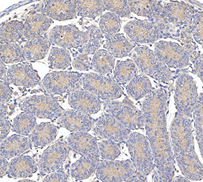 Immunohistochemical analysis of formalin fixed paraffin embedded mouse testicles tissue with F3225 at 1:10 dilution.