|
Wie zu zitieren 1. Für Zitate im Text (Materialien & Methoden): 2. Für die Tabelle der Schlüsselressourcen: |
||
|
Gebührenfrei: (877) 796-6397 -- Nur USA und Kanada -- |
Fax: +1-832-582-8590 Bestellungen: +1-832-582-8158 |
Technischer Support: +1-832-582-8158 Ext:3 Bitte geben Sie Ihre Bestellnummer in der E-Mail an. Wir bemühen uns, alle E-Mail-Anfragen innerhalb eines Werktages zu beantworten. |
Biologische Beschreibung
| Spezifität | Melanoma Associated Antigen 100+ / 7 kDa Antibody [G24P23] weist endogene Spiegel des gesamten Melanoma Associated Antigen 100+ / 7 kDa Proteins nach. |
|---|---|
| Hintergrund | Melanoma Associated Antigen 100+ / 7 kDa (MAGE)-Proteine sind tumorassoziierte Antigene, die typischerweise in Keimzellen des Hodens in normalen erwachsenen Geweben vorkommen, aber in verschiedenen Krebsarten, einschließlich Melanom, Gliom, Lunge, Blase und multiplem Myelom, aberrativ exprimiert werden. Die Bezeichnung „100+/7 kDa“ bezieht sich auf Varianten oder Isoformen mit Molekulargewichten um 100 kDa und 7 kDa. Strukturell teilen MAGE-Proteine eine konservierte ~200 Aminosäuren umfassende MAGE-Homologiedomäne (MHD), die aus zwei geflügelten Helixdomänen besteht, die eine tiefe Spalte für die Peptid- oder Proteinbindung bilden, flankiert von variablen N- und C-terminalen Regionen. Funktionell können MAGE-Proteine als onkogene Treiber wirken, indem sie mit RING-type E3 ubiquitin ligases interagieren und Proteindegradationswege modulieren, die Apoptose, Proliferation und Metabolismus regulieren; zum Beispiel fördert MAGE-A3 das Tumorüberleben durch Stimulierung des TRIM28-vermittelten p53-Abbaus, während MAGE-A4 über verschiedene Proteinpartner Apoptose induzieren kann. Ihre tumorspezifische Expression, strukturelle Plastizität und onkogenen Rollen machen MAGEs sowohl zu Biomarkern als auch zu potenziellen therapeutischen Zielen in der Krebsimmuntherapie. |
Nutzungsinformationen
| Anwendung | IHC, FCM | Verdünnung |
|
||
|---|---|---|---|---|---|
| Reaktivität | Human | ||||
| Quelle | Mouse Monoclonal Antibody | MW | |||
| Lagerpuffer | PBS, pH 7.2+50% Glycerol+0.05% BSA+0.01% NaN3 | Lagerung (Ab dem Datum des Erhalts) |
-20°C (avoid freeze-thaw cycles), 2 years | ||
| IHC |
Experimental Protocol:
Deparaffinization/Rehydration
1. Deparaffinize/hydrate sections:
2. Incubate sections in three washes of xylene for 5 min each.
3. Incubate sections in two washes of 100% ethanol for 10 min each.
4. Incubate sections in two washes of 95% ethanol for 10 min each.
5. Wash sections two times in dH2O for 5 min each.
6.Antigen retrieval: For Citrate: Heat slides in a microwave submersed in 1X citrate unmasking solution until boiling is initiated; continue with 10 min at a sub-boiling temperature (95°-98°C). Cool slides on bench top for 30 min.
Staining
1. Wash sections in dH2O three times for 5 min each.
2. Incubate sections in 3% hydrogen peroxide for 10 min.
3. Wash sections in dH2O two times for 5 min each.
4. Wash sections in wash buffer for 5 min.
5. Block each section with 100–400 µl of blocking solution for 1 hr at room temperature.
6. Remove blocking solution and add 100–400 µl primary antibody diluent in to each section. Incubate overnight at 4°C.
7. Remove antibody solution and wash sections with wash buffer three times for 5 min each.
8. Cover section with 1–3 drops HRPas needed. Incubate in a humidified chamber for 30 min at room temperature.
9. Wash sections three times with wash buffer for 5 min each.
10. Add DAB Chromogen Concentrate to DAB Diluent and mix well before use.
11. Apply 100–400 µl DAB to each section and monitor closely. 1–10 min generally provides an acceptable staining intensity.
12. Immerse slides in dH2O.
13. If desired, counterstain sections with hematoxylin.
14. Wash sections in dH2O two times for 5 min each.
15. Dehydrate sections: Incubate sections in 95% ethanol two times for 10 sec each; Repeat in 100% ethanol, incubating sections two times for 10 sec each; Repeat in xylene, incubating sections two times for 10 sec each.
16. Mount sections with coverslips and mounting medium.
|
Referenzen
|
Anwendungsdaten
IHC
Validiert von Selleck
-
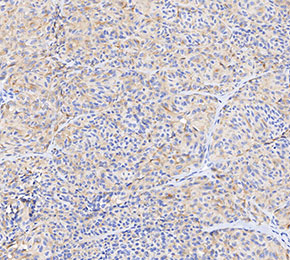 Immunohistochemical analysis of formalin fixed paraffin embedded human melanoma tissue with F3175 at 1:20 dilution.