|
Wie zu zitieren 1. Für Zitate im Text (Materialien & Methoden): 2. Für die Tabelle der Schlüsselressourcen: |
||
|
Gebührenfrei: (877) 796-6397 -- Nur USA und Kanada -- |
Fax: +1-832-582-8590 Bestellungen: +1-832-582-8158 |
Technischer Support: +1-832-582-8158 Ext:3 Bitte geben Sie Ihre Bestellnummer in der E-Mail an. Wir bemühen uns, alle E-Mail-Anfragen innerhalb eines Werktages zu beantworten. |
Biologische Beschreibung
| Spezifität | Perforin Antibody [P20D17] erkennt endogene Spiegel des gesamten Perforin-Proteins. |
|---|---|
| Hintergrund | Perforin ist ein porenformendes Glykoprotein, das hauptsächlich von zytotoxischen Lymphozyten wie natürlichen Killerzellen (NK-Zellen) und zytotoxischen T-Lymphozyten (CTLs) produziert wird. Es gehört zur MACPF-Familie (Membranangriffskomplex/Perforin) und besteht aus für seine Funktion kritischen Domänen: der MACPF-Domäne, die für die Porenformation verantwortlich ist, einer kalziumabhängigen C2-Domäne, die die Membranbindung vermittelt, und einer EGF-ähnlichen Domäne, die strukturelle Flexibilität bietet. Einmal in die immunologische Synapse sezerniert, die zwischen der zytotoxischen Zelle und der Zielzelle (virusinfiziert oder maligne) gebildet wird, oligomerisiert Perforin und insertiert in die Zielzellmembran, wobei transmembranäre Poren mit einem Durchmesser von etwa 20 nm gebildet werden. Diese Poren ermöglichen den Eintritt pro-apoptotischer Granzyme in das Zytosol, die die Apoptose der Zielzelle auslösen. Schlüsselreste wie R298 sind wichtig für die Oligomerisierung und Porenformation. Perforin-vermittelte Porenformation ist essenziell für die Immunüberwachung und Eliminierung abnormaler Zellen, und Defizite oder Mutationen in Perforin verursachen schwere Immundefizienzstörungen wie die familiäre hämophagozytische Lymphohistiozytose (FHL). Perforin spielt auch eine Rolle in der Immunregulation und ist beteiligt, wenn die zytotoxische Funktion bei verschiedenen Krankheiten gestört oder fehlreguliert ist. |
Nutzungsinformationen
| Anwendung | IHC | Verdünnung |
|
||
|---|---|---|---|---|---|
| Reaktivität | Human | ||||
| Quelle | Mouse Monoclonal Antibody | MW | 70 kDa | ||
| Lagerpuffer | PBS, pH 7.2+50% Glycerol+0.05% BSA+0.01% NaN3 | Lagerung (Ab dem Datum des Erhalts) |
-20°C (avoid freeze-thaw cycles), 2 years | ||
| IHC |
Experimental Protocol:
Deparaffinization/Rehydration
1. Deparaffinize/hydrate sections:
2. Incubate sections in three washes of xylene for 5 min each.
3. Incubate sections in two washes of 100% ethanol for 10 min each.
4. Incubate sections in two washes of 95% ethanol for 10 min each.
5. Wash sections two times in dH2O for 5 min each.
6.Antigen retrieval: For Citrate: Heat slides in a microwave submersed in 1X citrate unmasking solution until boiling is initiated; continue with 10 min at a sub-boiling temperature (95°-98°C). Cool slides on bench top for 30 min.
Staining
1. Wash sections in dH2O three times for 5 min each.
2. Incubate sections in 3% hydrogen peroxide for 10 min.
3. Wash sections in dH2O two times for 5 min each.
4. Wash sections in wash buffer for 5 min.
5. Block each section with 100–400 µl of blocking solution for 1 hr at room temperature.
6. Remove blocking solution and add 100–400 µl primary antibody diluent in to each section. Incubate overnight at 4°C.
7. Remove antibody solution and wash sections with wash buffer three times for 5 min each.
8. Cover section with 1–3 drops HRPas needed. Incubate in a humidified chamber for 30 min at room temperature.
9. Wash sections three times with wash buffer for 5 min each.
10. Add DAB Chromogen Concentrate to DAB Diluent and mix well before use.
11. Apply 100–400 µl DAB to each section and monitor closely. 1–10 min generally provides an acceptable staining intensity.
12. Immerse slides in dH2O.
13. If desired, counterstain sections with hematoxylin.
14. Wash sections in dH2O two times for 5 min each.
15. Dehydrate sections: Incubate sections in 95% ethanol two times for 10 sec each; Repeat in 100% ethanol, incubating sections two times for 10 sec each; Repeat in xylene, incubating sections two times for 10 sec each.
16. Mount sections with coverslips and mounting medium.
|
Referenzen
|
Anwendungsdaten
IHC
Validiert von Selleck
-
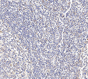 Immunohistochemical analysis of formalin fixed paraffin embedded human tonsils tissue with F2444 at 1:100 dilution.