|
Wie zu zitieren 1. Für Zitate im Text (Materialien & Methoden): 2. Für die Tabelle der Schlüsselressourcen: |
||
|
Gebührenfrei: (877) 796-6397 -- Nur USA und Kanada -- |
Fax: +1-832-582-8590 Bestellungen: +1-832-582-8158 |
Technischer Support: +1-832-582-8158 Ext:3 Bitte geben Sie Ihre Bestellnummer in der E-Mail an. Wir bemühen uns, alle E-Mail-Anfragen innerhalb eines Werktages zu beantworten. |
Biologische Beschreibung
| Spezifität | Phospho-Akt1 (Ser129) Antibody [K23C4] detektiert endogene Spiegel des gesamten Akt1-Proteins nur, wenn es an Ser129 phosphoryliert ist. |
|---|---|
| Hintergrund | Phospho-Akt1 (Ser129) ist eine spezifische posttranslationale Modifikation von Akt1 (RAC-alpha Serin/Threonin-Proteinkinase, PKBα), die innerhalb der flexiblen Linkerregion zwischen der Pleckstrin-Homologie (PH)-Domäne und der Kinasedomäne dieses 480 Aminosäuren umfassenden Proteins auftritt. Akt1 besteht aus einer N-terminalen PH-Domäne für die PIP3-vermittelte Membranrekrutierung, einer katalytischen Kinasedomäne mit wichtigen regulatorischen Phosphorylierungsstellen an Thr308 (Aktivierungsschleife) und Ser473 (hydrophobes Motiv) sowie einem C-terminalen regulatorischen Schwanz. Die Phosphorylierung von Ser129 durch die Proteinkinase CK2 erzeugt ein Konsensus-S-x-x-D/E-Motiv, das eine nachfolgende Modifikation an Ser126 vorbereitet und die Gesamtkonformation von Akt1 stabilisiert. Diese Modifikation hyperaktiviert Akt1 über die Effekte der kanonischen PDK1/mTORC2-Phosphorylierung hinaus, indem sie dessen Assoziation mit dem HSP90-Chaperonkomplex stärkt, der Thr308 vor PP2A-vermittelter Dephosphorylierung schützt und dadurch die Akt1-Kinaseaktivität aufrechterhält. Zusätzlich verbessert Phospho-Ser129 die β-Catenin/TCF-Transkriptionsaktivität, entweder direkt oder durch Stabilisierung von Wnt-Signalgebungskomponenten, und fixiert Akt1 in einer erweiterten aktiven Konformation, die eine isoformenspezifische Substratselektion ermöglicht, wie die bevorzugte Phosphorylierung von Palladin, ein Prozess, der bei Akt2 aufgrund des Fehlens der äquivalenten Ser131-Stelle nicht auftritt. Diese Modifikation treibt die Umgestaltung des Zytoskeletts, das Zellüberleben durch Hemmung von Bad und FoxO, die Proliferation durch Förderung der Cyclin-D1-Stabilität und p27/p21-Sequestrierung sowie die mTORC1-Aktivierung über die TSC2-Phosphorylierung voran, wobei die hierarchische Phosphorylierung eine robuste Signalverstärkung bei Wachstumsfaktorstimulation gewährleistet. Bei Krankheiten erhöhen erhöhte Phospho-Ser129-Spiegel das Überleben und die Metastasierung von Krebszellen durch Verstärkung der PI3K/Akt-onkogenen Signalgebung (insbesondere bei Brust- und Prostatakrebs), tragen zur Chemoresistenz durch anhaltenden HSP90-Schutz bei und sind an Stoffwechselstörungen durch die Störung der Glukosehomöostase durch Hyperaktivierung von Akt1 beteiligt. |
Nutzungsinformationen
| Anwendung | WB | Verdünnung |
|
||
|---|---|---|---|---|---|
| Reaktivität | Human | ||||
| Quelle | Rabbit Monoclonal Antibody | MW | 55 kDa | ||
| Lagerpuffer | PBS, pH 7.2+50% Glycerol+0.05% BSA+0.01% NaN3 | Lagerung (Ab dem Datum des Erhalts) |
-20°C (avoid freeze-thaw cycles), 2 years | ||
| WB |
Experimental Protocol:
Sample preparation
1. Tissue: Lyse the tissue sample by adding an appropriate volume of ice-cold RIPA/NP-40 Lysis Buffer (containing Protease Inhibitor Cocktail, Phosphatase Inhibitor Cocktail),and homogenize the tissue at a low temperature. 2. Adherent cell: Aspirate the culture medium and wash the cells with ice-cold PBS twice. Lyse the cells by adding an appropriate volume of RIPA/NP-40 Lysis Buffer (containing Protease Inhibitor Cocktail, Phosphatase Inhibitor Cocktail) and put the sample on ice for 5 min. 3. Suspension cell: Transfer the culture medium to a pre-cooled centrifuge tube. Centrifuge and aspirate the supernatant. Wash the cells with ice-cold PBS twice. Lyse the cells by adding an appropriate volume of RIPA/NP-40 Lysis Buffer (containing Protease Inhibitor Cocktail, Phosphatase Inhibitor Cocktail) and put the sample on ice for 5 min. 4. Place the lysate into a pre-cooled microcentrifuge tube. Centrifuge at 4°C for 15 min. Collect the supernatant;
5. Remove a small volume of lysate to determine the protein concentration;
6. Combine the lysate with protein loading buffer. Boil 20 µL sample under 95-100°C for 5 min. Centrifuge for 5 min after cool down on ice.
Electrophoretic separation
1. According to the concentration of extracted protein, load appropriate amount of protein sample and marker onto SDS-PAGE gels for electrophoresis. Recommended separating gel (lower gel) concentration: 10%. Reference Table for Selecting SDS-PAGE Separation Gel Concentrations 2. Power up 80V for 30 minutes. Then the power supply is adjusted (110 V~150 V), the Marker is observed, and the electrophoresis can be stopped when the indicator band of the predyed protein Marker where the protein is located is properly separated. (Note that the current should not be too large when electrophoresis, too large current (more than 150 mA) will cause the temperature to rise, affecting the result of running glue. If high currents cannot be avoided, an ice bath can be used to cool the bath.)
Transfer membrane
1. Take out the converter, soak the clip and consumables in the pre-cooled converter;
2. Activate PVDF membrane with methanol for 1 min and rinse with transfer buffer;
3. Install it in the order of "black edge of clip - sponge - filter paper - filter paper - glue -PVDF membrane - filter paper - filter paper - sponge - white edge of clip"; 4. The protein was electrotransferred to PVDF membrane. ( 0.45 µm PVDF membrane is recommended ) Reference Table for Selecting PVDF Membrane Pore Size Specifications Recommended conditions for wet transfer: 200 mA, 120 min. ( Note that the transfer conditions can be adjusted according to the protein size. For high-molecular-weight proteins, a higher current and longer transfer time are recommended. However, ensure that the transfer tank remains at a low temperature to prevent gel melting.)
Block
1. After electrotransfer, wash the film with TBST at room temperature for 5 minutes;
2. Incubate the film in the blocking solution ( recommending 5% BSA solution)
for 1 hour at room temperature;
3. Wash the film with TBST for 3 times, 5 minutes each time.
Antibody incubation
1. Use 5% skim milk powder to prepare the primary antibody working liquid (recommended dilution ratio for primary antibody 1:1000), gently shake and incubate with the film at 4°C overnight; 2. Wash the film with TBST 3 times, 5 minutes each time;
3. Add the secondary antibody to the blocking solution and incubate with the film gently at room temperature for 1 hour;
4. After incubation, wash the film with TBST 3 times for 5 minutes each time.
Antibody staining
1. Add the prepared ECL luminescent substrate (or select other color developing substrate according to the second antibody) and mix evenly;
2. Incubate with the film for 1 minute, remove excess substrate (keep the film moist), wrap with plastic film, and expose in the imaging system.
|
Referenzen
|
Anwendungsdaten
WB
Validiert von Selleck
-
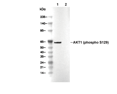 Lane 1: MCF-7, Lane 2: MCF-7 (Alkaline Phosphatase treated)