|
Wie zu zitieren 1. Für Zitate im Text (Materialien & Methoden): 2. Für die Tabelle der Schlüsselressourcen: |
||
|
Gebührenfrei: (877) 796-6397 -- Nur USA und Kanada -- |
Fax: +1-832-582-8590 Bestellungen: +1-832-582-8158 |
Technischer Support: +1-832-582-8158 Ext:3 Bitte geben Sie Ihre Bestellnummer in der E-Mail an. Wir bemühen uns, alle E-Mail-Anfragen innerhalb eines Werktages zu beantworten. |
Biologische Beschreibung
| Spezifität | Phospho-IκBα (Ser32/36) Antibody [F24N7] weist endogene IκBα-Spiegel nur nach, wenn diese an Ser32/36 phosphoryliert sind. |
|---|---|
| Hintergrund | Der IκB-Kinase (IKK)-Komplex dient als zentraler Signalgeber für die Aktivierung des NF-κB-Signalwegs. Dieser Komplex besteht aus zwei katalytischen Untereinheiten, IKKα und IKKβ, die Serin/Threonin-Kinasen sind, und einer regulatorischen Untereinheit, bekannt als NEMO (NF-κB essential modulator), auch als IKKγ bezeichnet. Der IKK-Komplex integriert eine Vielzahl von vorgelagerten Signalen, die NF-κB aktivieren, was zur Phosphorylierung von IκB-Proteinen, NF-κB-Untereinheiten und anderen zellulären Zielen führt. In unstimulierten Zellen werden NF-κB-Transkriptionsfaktoren im Zytoplasma durch Bindung an inhibitorische IκB-Proteine wie IκBα, IκBβ und IκBε oder an die Vorläuferproteine p100 und p105 in einem inaktiven Zustand gehalten. Diese inhibitorischen Proteine verhindern, dass NF-κB in den Zellkern gelangt und die Gentranskription initiiert. Nach Stimulation werden IκB-Proteine an spezifischen Serinresten – insbesondere Ser32 und Ser36 im Falle von IκBα – durch den aktivierten IKK-Komplex phosphoryliert. Diese Phosphorylierung markiert IκB für die Erkennung durch den SCFβTrCP E3 Ubiquitinligase-Komplex, was zu seiner Ubiquitinierung und anschließendem Abbau über das Proteasom führt. Der Abbau von IκB setzt NF-κB-Dimere frei, die dann in den Zellkern translozieren, wo sie die Transkription von Zielgenen steuern. Eine Vielzahl externer Signale, einschließlich entzündlicher Zytokine, Wachstumsfaktoren und Chemokine, kann diese Kaskade auslösen, was letztendlich zur Phosphorylierung von IκBα und Aktivierung der NF-κB-abhängigen Genexpression führt. |
Nutzungsinformationen
| Anwendung | WB | Verdünnung |
|
||
|---|---|---|---|---|---|
| Reaktivität | Human, Mouse, Rat, Monkey | ||||
| Quelle | Mouse Monoclonal Antibody | MW | 40 kDa | ||
| Lagerpuffer | PBS, pH 7.2+50% Glycerol+0.05% BSA+0.01% NaN3 | Lagerung (Ab dem Datum des Erhalts) |
-20°C (avoid freeze-thaw cycles), 2 years | ||
| WB |
Experimental Protocol:
Sample preparation
1. Tissue: Lyse the tissue sample by adding an appropriate volume of ice-cold RIPA/Nuclear Lysis Buffer (containing Protease Inhibitor Cocktail, Phosphatase Inhibitor Cocktail),and homogenize the tissue at a low temperature. 2. Adherent cell: Aspirate the culture medium and wash the cells with ice-cold PBS twice. Lyse the cells by adding an appropriate volume of RIPA/Nuclear Lysis Buffer (containing Protease Inhibitor Cocktail, Phosphatase Inhibitor Cocktail) and put the sample on ice for 5 min. 3. Suspension cell: Transfer the culture medium to a pre-cooled centrifuge tube. Centrifuge and aspirate the supernatant. Wash the cells with ice-cold PBS twice. Lyse the cells by adding an appropriate volume of RIPA/Nuclear Lysis Buffer (containing Protease Inhibitor Cocktail, Phosphatase Inhibitor Cocktail) and put the sample on ice for 5 min. 4. Place the lysate into a pre-cooled microcentrifuge tube. Centrifuge at 4°C for 15 min. Collect the supernatant;
5. Remove a small volume of lysate to determine the protein concentration;
6. Combine the lysate with protein loading buffer. Boil 20 µL sample under 95-100°C for 5 min. Centrifuge for 5 min after cool down on ice.
Electrophoretic separation
1. According to the concentration of extracted protein, load appropriate amount of protein sample and marker onto SDS-PAGE gels for electrophoresis. Recommended separating gel (lower gel) concentration: 10%. Reference Table for Selecting SDS-PAGE Separation Gel Concentrations 2. Power up 80V for 30 minutes. Then the power supply is adjusted (110 V~150 V), the Marker is observed, and the electrophoresis can be stopped when the indicator band of the predyed protein Marker where the protein is located is properly separated. (Note that the current should not be too large when electrophoresis, too large current (more than 150 mA) will cause the temperature to rise, affecting the result of running glue. If high currents cannot be avoided, an ice bath can be used to cool the bath.)
Transfer membrane
1. Take out the converter, soak the clip and consumables in the pre-cooled converter;
2. Activate PVDF membrane with methanol for 1 min and rinse with transfer buffer;
3. Install it in the order of "black edge of clip - sponge - filter paper - filter paper - glue -PVDF membrane - filter paper - filter paper - sponge - white edge of clip"; 4. The protein was electrotransferred to PVDF membrane. ( 0.45 µm PVDF membrane is recommended ) Reference Table for Selecting PVDF Membrane Pore Size Specifications Recommended conditions for wet transfer: 200 mA, 60 min. ( Note that the transfer conditions can be adjusted according to the protein size. For high-molecular-weight proteins, a higher current and longer transfer time are recommended. However, ensure that the transfer tank remains at a low temperature to prevent gel melting.)
Block
1. After electrotransfer, wash the film with TBST at room temperature for 5 minutes;
2. Incubate the film in the blocking solution ( recommending 5% BSA solution)
for 1 hour at room temperature;
3. Wash the film with TBST for 3 times, 5 minutes each time.
Antibody incubation
1. Use 5% skim milk powder to prepare the primary antibody working liquid (recommended dilution ratio for primary antibody 1:1000), gently shake and incubate with the film at 4°C overnight; 2. Wash the film with TBST 3 times, 5 minutes each time;
3. Add the secondary antibody to the blocking solution and incubate with the film gently at room temperature for 1 hour;
4. After incubation, wash the film with TBST 3 times for 5 minutes each time.
Antibody staining
1. Add the prepared ECL luminescent substrate (or select other color developing substrate according to the second antibody) and mix evenly;
2. Incubate with the film for 1 minute, remove excess substrate (keep the film moist), wrap with plastic film, and expose in the imaging system. (Exposure time of at least 150s is recommended)
|
Referenzen
|
Anwendungsdaten
WB
Validiert von Selleck
-
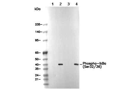 Lane 1: Hela, Lane 2: Hela (IFNa, 20 ng/mL, 5 min), Lane 3: 3T3, Lane 4: 3T3 (IFNa, 20 ng/mL, 5 min)