|
Wie zu zitieren 1. Für Zitate im Text (Materialien & Methoden): 2. Für die Tabelle der Schlüsselressourcen: |
||
|
Gebührenfrei: (877) 796-6397 -- Nur USA und Kanada -- |
Fax: +1-832-582-8590 Bestellungen: +1-832-582-8158 |
Technischer Support: +1-832-582-8158 Ext:3 Bitte geben Sie Ihre Bestellnummer in der E-Mail an. Wir bemühen uns, alle E-Mail-Anfragen innerhalb eines Werktages zu beantworten. |
Biologische Beschreibung
| Spezifität | Phospho-RIP3 (Ser232) Antibody [E19K17] erkennt endogene Spiegel des gesamten RIP3-Proteins nur, wenn es an Ser232 phosphoryliert ist |
|---|---|
| Hintergrund | Die Rezeptor-interagierende Protein (RIP)-Familie der Serin/Threonin-Kinasen – einschließlich RIP1, RIP2, RIP3 und RIP4 – spielt eine entscheidende Rolle bei der Modulation zellulärer Stressreaktionen. Diese Kinasen sind an der Initiierung von Pro-Survival- und Entzündungssignalen, hauptsächlich durch NF-κB-Aktivierung, sowie an der Förderung von Zelltodwegen wie Apoptosis beteiligt. Unter ihnen ist die Rezeptor-interagierende Proteinkinase 3 (RIPK3 oder RIP3) ein zentraler Akteur bei der Nekroptose, einer regulierten Form des nekrotischen Zelltods. RIP3 ist an Signalkaskaden beteiligt, die durch den Tumornekrosefaktor (TNF) initiiert werden, wo es sich mit RIP1 und dem TNF-Rezeptorkomplex assoziiert, um sowohl die NF-κB-Aktivierung als auch Apoptosis zu vermitteln. Ein Schlüsselereignis bei der Nekroptose ist die Interaktion zwischen RIP1 und RIP3, die die Grundlage des Nekrosoms bildet – eines Multiproteinkomplexes, der die programmierte Nekrose erleichtert. Dieser nekroseartige Zelltod wird typischerweise durch TNF-Signale in Anwesenheit von Caspase-Inhibition ausgelöst. Beim Menschen ist die Phosphorylierung von RIP3 an Serin 227 entscheidend für seine Bindung an das Mixed-Lineage-Kinase-Domänen-ähnliche Protein (MLKL), wodurch die Nekrosom-Assemblierung ermöglicht wird. Bei Mäusen induziert die TNF-Stimulation die Phosphorylierung an Threonin 231 und Serin 232 an RIP3, was für seine Interaktion mit Maus-MLKL notwendig ist. Bemerkenswerterweise entspricht Ser-232 in murine RIP3 Ser-227 im menschlichen Protein. Zusätzlich tragen RIP1 und RIP3 zur Oligomerisierung des Nekrosoms bei, was die Bildung von Amyloid-ähnlichen Signalstrukturen fördert, die für die Ausführung der Nekroptose unerlässlich sind. |
Nutzungsinformationen
| Anwendung | WB, ELISA | Verdünnung |
|
||
|---|---|---|---|---|---|
| Reaktivität | Mouse | ||||
| Quelle | Rabbit Monoclonal Antibody | MW | 53 kDa | ||
| Lagerpuffer | PBS, pH 7.2+50% Glycerol+0.05% BSA+0.01% NaN3 | Lagerung (Ab dem Datum des Erhalts) |
-20°C (avoid freeze-thaw cycles), 2 years | ||
| WB |
Experimental Protocol:
Sample preparation
1. Tissue: Lyse the tissue sample by adding an appropriate volume of ice-cold RIPA/Nuclear Lysis Buffer (containing Protease Inhibitor Cocktail, Phosphatase Inhibitor Cocktail),and homogenize the tissue at a low temperature. 2. Adherent cell: Aspirate the culture medium and wash the cells with ice-cold PBS twice. Lyse the cells by adding an appropriate volume of RIPA/Nuclear Lysis Buffer (containing Protease Inhibitor Cocktail, Phosphatase Inhibitor Cocktail) and put the sample on ice for 5 min. 3. Suspension cell: Transfer the culture medium to a pre-cooled centrifuge tube. Centrifuge and aspirate the supernatant. Wash the cells with ice-cold PBS twice. Lyse the cells by adding an appropriate volume of RIPA/Nuclear Lysis Buffer (containing Protease Inhibitor Cocktail, Phosphatase Inhibitor Cocktail) and put the sample on ice for 5 min. 4. Place the lysate into a pre-cooled microcentrifuge tube. Centrifuge at 4°C for 15 min. Collect the supernatant;
5. Remove a small volume of lysate to determine the protein concentration;
6. Combine the lysate with protein loading buffer. Boil 20 µL sample under 95-100°C for 5 min. Centrifuge for 5 min after cool down on ice.
Electrophoretic separation
1. According to the concentration of extracted protein, load appropriate amount of protein sample and marker onto SDS-PAGE gels for electrophoresis. Recommended separating gel (lower gel) concentration: 10%. Reference Table for Selecting SDS-PAGE Separation Gel Concentrations 2. Power up 80V for 30 minutes. Then the power supply is adjusted (110 V~150 V), the Marker is observed, and the electrophoresis can be stopped when the indicator band of the predyed protein Marker where the protein is located is properly separated. (Note that the current should not be too large when electrophoresis, too large current (more than 150 mA) will cause the temperature to rise, affecting the result of running glue. If high currents cannot be avoided, an ice bath can be used to cool the bath.)
Transfer membrane
1. Take out the converter, soak the clip and consumables in the pre-cooled converter;
2. Activate PVDF membrane with methanol for 1 min and rinse with transfer buffer;
3. Install it in the order of "black edge of clip - sponge - filter paper - filter paper - glue -PVDF membrane - filter paper - filter paper - sponge - white edge of clip"; 4. The protein was electrotransferred to PVDF membrane. ( 0.45 µm PVDF membrane is recommended ) Reference Table for Selecting PVDF Membrane Pore Size Specifications Recommended conditions for wet transfer: 200 mA, 120 min. ( Note that the transfer conditions can be adjusted according to the protein size. For high-molecular-weight proteins, a higher current and longer transfer time are recommended. However, ensure that the transfer tank remains at a low temperature to prevent gel melting.)
Block
1. After electrotransfer, wash the film with TBST at room temperature for 5 minutes;
2. Incubate the film in the blocking solution ( recommending 5% BSA solution)
for 1 hour at room temperature;
3. Wash the film with TBST for 3 times, 5 minutes each time.
Antibody incubation
1. Use 5% skim milk powder to prepare the primary antibody working liquid (recommended dilution ratio for primary antibody 1:1000), gently shake and incubate with the film at 4°C overnight; 2. Wash the film with TBST 3 times, 5 minutes each time;
3. Add the secondary antibody to the blocking solution and incubate with the film gently at room temperature for 1 hour;
4. After incubation, wash the film with TBST 3 times for 5 minutes each time.
Antibody staining
1. Add the prepared ECL luminescent substrate (or select other color developing substrate according to the second antibody) and mix evenly;
2. Incubate with the film for 1 minute, remove excess substrate (keep the film moist), wrap with plastic film, and expose in the imaging system.
|
Referenzen
|
Anwendungsdaten
WB
Validiert von Selleck
-
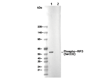 Lane 1: L-929, Lane 2: L-929 (phosphatase-treated)