|
Wie zu zitieren 1. Für Zitate im Text (Materialien & Methoden): 2. Für die Tabelle der Schlüsselressourcen: |
||
|
Gebührenfrei: (877) 796-6397 -- Nur USA und Kanada -- |
Fax: +1-832-582-8590 Bestellungen: +1-832-582-8158 |
Technischer Support: +1-832-582-8158 Ext:3 Bitte geben Sie Ihre Bestellnummer in der E-Mail an. Wir bemühen uns, alle E-Mail-Anfragen innerhalb eines Werktages zu beantworten. |
Biologische Beschreibung
| Spezifität | Phospho-Vimentin (Ser39) Antibody [B14E20] erkennt endogene Spiegel des gesamten Vimentin-Proteins nur, wenn es an Ser39 phosphoryliert ist. |
|---|---|
| Hintergrund | Phospho-Vimentin (Ser39) bezieht sich auf das Vimentin-Intermediärfilamentprotein, das an Serin 39 phosphoryliert ist, eine Modifikation, die eine entscheidende Rolle bei der Regulierung der Filamentdynamik und der zellulären Architektur spielt. Vimentin ist ein Intermediärfilamentprotein vom Typ III, das überwiegend in mesenchymalen Zellen exprimiert wird und als Hauptbestandteil des Zytoskeletts dient, das mechanische Stabilität bietet, die zelluläre Integrität aufrechterhält und den intrazellulären Transport unterstützt. Vimentin lagert sich hierarchisch zusammen: Lösliche Coiled-Coil-Dimere richten sich antiparallel aus, um A11-Tetramere zu bilden, die sich dann lateral und longitudinal zu reifen Filamenten mit 11 nm Durchmesser verbinden. Die Kopfflüssigkeit, in der Ser39 lokalisiert ist, ist essentiell für die Nukleation der Filamentzusammenlagerung und dient als Hotspot für die regulatorische Phosphorylierung. Die Phosphorylierung an Ser39 induziert Konformationsänderungen, die die Filamentwechselwirkungen schwächen und die Disassemblierung oder Reorganisation fördern, ein Prozess, der während der Mitose, der Zellmigration und als Reaktion auf mechanischen Stress entscheidend ist. Diese dynamische Umgestaltung ermöglicht es Vimentin, als Signalgerüst zu fungieren und Änderungen in der Zellform, der Motilität und der intrazellulären Signalgebung zu koordinieren. Der reversible Montage- und Demontagezyklus, der streng durch Phosphorylierungsereignisse wie an Ser39 reguliert wird, ermöglicht es Zellen, ihre mechanischen Eigenschaften schnell an physiologische Anforderungen anzupassen. Eine Dysregulation der Vimentin-Phosphorylierung ist an Krebsmetastasierung, Fibrose und anderen Pathologien beteiligt. |
Nutzungsinformationen
| Anwendung | WB | Verdünnung |
|
||
|---|---|---|---|---|---|
| Reaktivität | Human | ||||
| Quelle | Rabbit Monoclonal Antibody | MW | 54 kDa | ||
| Lagerpuffer | PBS, pH 7.2+50% Glycerol+0.05% BSA+0.01% NaN3 | Lagerung (Ab dem Datum des Erhalts) |
-20°C (avoid freeze-thaw cycles), 2 years | ||
| WB |
Experimental Protocol:
Sample preparation
1. Tissue: Lyse the tissue sample by adding an appropriate volume of ice-cold RIPA/NP-40 Lysis Buffer (containing Protease Inhibitor Cocktail, Phosphatase Inhibitor Cocktail),and homogenize the tissue at a low temperature. 2. Adherent cell: Aspirate the culture medium and wash the cells with ice-cold PBS twice. Lyse the cells by adding an appropriate volume of RIPA/NP-40 Lysis Buffer (containing Protease Inhibitor Cocktail, Phosphatase Inhibitor Cocktail) and put the sample on ice for 5 min. 3. Suspension cell: Transfer the culture medium to a pre-cooled centrifuge tube. Centrifuge and aspirate the supernatant. Wash the cells with ice-cold PBS twice. Lyse the cells by adding an appropriate volume of RIPA/NP-40 Lysis Buffer (containing Protease Inhibitor Cocktail, Phosphatase Inhibitor Cocktail) and put the sample on ice for 5 min. 4. Place the lysate into a pre-cooled microcentrifuge tube. Centrifuge at 4°C for 15 min. Collect the supernatant;
5. Remove a small volume of lysate to determine the protein concentration;
6. Combine the lysate with protein loading buffer. Boil 20 µL sample under 95-100°C for 5 min. Centrifuge for 5 min after cool down on ice.
Electrophoretic separation
1. According to the concentration of extracted protein, load appropriate amount of protein sample and marker onto SDS-PAGE gels for electrophoresis. Recommended separating gel (lower gel) concentration: 10%. Reference Table for Selecting SDS-PAGE Separation Gel Concentrations 2. Power up 80V for 30 minutes. Then the power supply is adjusted (110 V~150 V), the Marker is observed, and the electrophoresis can be stopped when the indicator band of the predyed protein Marker where the protein is located is properly separated. (Note that the current should not be too large when electrophoresis, too large current (more than 150 mA) will cause the temperature to rise, affecting the result of running glue. If high currents cannot be avoided, an ice bath can be used to cool the bath.)
Transfer membrane
1. Take out the converter, soak the clip and consumables in the pre-cooled converter;
2. Activate PVDF membrane with methanol for 1 min and rinse with transfer buffer;
3. Install it in the order of "black edge of clip - sponge - filter paper - filter paper - glue -PVDF membrane - filter paper - filter paper - sponge - white edge of clip"; 4. The protein was electrotransferred to PVDF membrane. ( 0.45 µm PVDF membrane is recommended ) Reference Table for Selecting PVDF Membrane Pore Size Specifications Recommended conditions for wet transfer: 200 mA, 120 min. ( Note that the transfer conditions can be adjusted according to the protein size. For high-molecular-weight proteins, a higher current and longer transfer time are recommended. However, ensure that the transfer tank remains at a low temperature to prevent gel melting.)
Block
1. After electrotransfer, wash the film with TBST at room temperature for 5 minutes;
2. Incubate the film in the blocking solution ( recommending 5% BSA solution)
for 1 hour at room temperature;
3. Wash the film with TBST for 3 times, 5 minutes each time.
Antibody incubation
1. Use 5% skim milk powder to prepare the primary antibody working liquid (recommended dilution ratio for primary antibody 1:1000), gently shake and incubate with the film at 4°C overnight; 2. Wash the film with TBST 3 times, 5 minutes each time;
3. Add the secondary antibody to the blocking solution and incubate with the film gently at room temperature for 1 hour;
4. After incubation, wash the film with TBST 3 times for 5 minutes each time.
Antibody staining
1. Add the prepared ECL luminescent substrate (or select other color developing substrate according to the second antibody) and mix evenly;
2. Incubate with the film for 1 minute, remove excess substrate (keep the film moist), wrap with plastic film, and expose in the imaging system.
|
Referenzen
|
Anwendungsdaten
WB
Validiert von Selleck
-
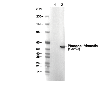 Lane 1: Hela, Lane 2: Hela (Calyculin A-treated)