|
Wie zu zitieren 1. Für Zitate im Text (Materialien & Methoden): 2. Für die Tabelle der Schlüsselressourcen: |
||
|
Gebührenfrei: (877) 796-6397 -- Nur USA und Kanada -- |
Fax: +1-832-582-8590 Bestellungen: +1-832-582-8158 |
Technischer Support: +1-832-582-8158 Ext:3 Bitte geben Sie Ihre Bestellnummer in der E-Mail an. Wir bemühen uns, alle E-Mail-Anfragen innerhalb eines Werktages zu beantworten. |
Biologische Beschreibung
| Spezifität | PI3 Kinase p110 δ Antibody [H17B15] erkennt endogene Spiegel des gesamten PI3 Kinase Class III δ-Proteins. |
|---|---|
| Hintergrund | Die Phosphoinositid-3-Kinase (PI3K) ist ein Enzym, das Phosphatidylinositol (PI), Phosphatidylinositol-4-phosphat (PIP) und Phosphatidylinositol-4,5-bisphosphat (PIP2) phosphoryliert, um Phosphatidylinositol-3,4,5-triphosphat zu erzeugen. Dieser Prozess wird durch Wachstumsfaktoren und Hormone aktiviert und führt zur Regulierung des Zellwachstums, der Zellzyklusprogression, der Zellmigration und des Überlebens. Das Enzym PTEN wirkt diesem Prozess entgegen, indem es Phosphatidylinositol-3,4,5-triphosphat dephosphoryliert, und Studien haben gezeigt, dass der Verlust der PTEN-Funktion zu einer konstitutiven Aktivierung des PI3K-Signalwegs bei verschiedenen menschlichen Krebsarten führt. PI3Ks bestehen aus einer katalytischen Untereinheit (p110) und einer regulatorischen Untereinheit, wobei verschiedene Isoformen der katalytischen Untereinheit, einschließlich p110α, p110β, p110γ und p110δ, existieren. Die regulatorischen Untereinheiten p85α und p85β interagieren mit p110α, p110β und p110δ. Während Gain-of-Function-Mutationen im PIK3CA-Gen, das die p110α-Isoform kodiert, häufig bei vielen menschlichen Krebsarten beobachtet werden, wurden keine somatischen Mutationen in Genen gefunden, die für p110β oder p110δ kodieren. Im Gegensatz zu den breit exprimierten p110α und p110β wird p110δ überwiegend in Leukozyten gefunden, was den p110δ-Signalweg zu einem Forschungsschwerpunkt bei Immunerkrankungen macht. Die p110δ-Isoform spielt eine Schlüsselrolle bei der Entwicklung und Progression bestimmter hämatologischer Malignome, wobei Studien mit p110δ-selektiven Inhibitoren und genetischer Inaktivierung in Mausmodellen ihre Beteiligung an Prozessen wie Zelldifferenzierung, Wachstum, Überleben, Motilität und Interaktion mit der Inositolphosphatase PTEN hervorheben. |
Nutzungsinformationen
| Anwendung | WB, IP | Verdünnung |
|
||||
|---|---|---|---|---|---|---|---|
| Reaktivität | Human, Mouse, Rat | ||||||
| Quelle | Rabbit Monoclonal Antibody | MW | 110 kDa | ||||
| Lagerpuffer | PBS, pH 7.2+50% Glycerol+0.05% BSA+0.01% NaN₃ | Lagerung (Ab dem Datum des Erhalts) |
-20°C (avoid freeze-thaw cycles), 2 years | ||||
| WB |
Experimental Protocol:
Sample preparation
1. Tissue: Lyse the tissue sample by adding an appropriate volume of ice-cold RIPA/Tris-HCL Lysis Buffer (containing Protease Inhibitor Cocktail),and homogenize the tissue at a low temperature or lyse it by sonication on ice, then incubate on ice for 30 minutes. 2. Adherent cell: Aspirate the culture medium and transfer the cells into an EP tube. Wash the cells with ice-cold PBS twice. Add an appropriate volume of RIPA/Tris-HCL Lysis Buffer (containing Protease Inhibitor Cocktail), sonicate to lyse the cells, and incubate on ice for 30 minutes. 3. Suspension cell: Transfer the culture medium to a pre-cooled centrifuge tube. Centrifuge and aspirate the supernatant. Wash the cells with ice-cold PBS twice.Add an appropriate volume of RIPA/Tris-HCL Lysis Buffer (containing Protease Inhibitor Cocktail), sonicate to lyse the cells, and incubate on ice for 30 minutes. 4. Place the lysate into a pre-cooled microcentrifuge tube. Centrifuge at 4°C for 15 min. Collect the supernatant;
5. Remove a small volume of lysate to determine the protein concentration;
6. Combine the lysate with protein loading buffer. Boil 20 µL sample under 95-100°C for 5 min. Centrifuge for 5 min after cool down on ice.
Electrophoretic separation
1. According to the concentration of extracted protein, load appropriate amount of protein sample and marker onto SDS-PAGE gels for electrophoresis. Recommended separating gel (lower gel) concentration: 5%. Reference Table for Selecting SDS-PAGE Separation Gel Concentrations 2. Power up 80V for 30 minutes. Then the power supply is adjusted (110 V~150 V), the Marker is observed, and the electrophoresis can be stopped when the indicator band of the predyed protein Marker where the protein is located is properly separated. (Note that the current should not be too large when electrophoresis, too large current (more than 150 mA) will cause the temperature to rise, affecting the result of running glue. If high currents cannot be avoided, an ice bath can be used to cool the bath.)
Transfer membrane
1. Take out the converter, soak the clip and consumables in the pre-cooled converter;
2. Activate PVDF membrane with methanol for 1 min and rinse with transfer buffer;
3. Install it in the order of "black edge of clip - sponge - filter paper - filter paper - glue -PVDF membrane - filter paper - filter paper - sponge - white edge of clip"; 4. The protein was electrotransferred to PVDF membrane. ( 0.45 µm PVDF membrane is recommended ) Reference Table for Selecting PVDF Membrane Pore Size Specifications Recommended conditions for wet transfer: 200 mA, 120 min. ( Note that the transfer conditions can be adjusted according to the protein size. For high-molecular-weight proteins, a higher current and longer transfer time are recommended. However, ensure that the transfer tank remains at a low temperature to prevent gel melting.)
Block
1. After electrotransfer, wash the film with TBST at room temperature for 5 minutes;
2. Incubate the film in the blocking solution for 1 hour at room temperature;
3. Wash the film with TBST for 3 times, 5 minutes each time.
Antibody incubation
1. Use 5% skim milk powder to prepare the primary antibody working liquid (recommended dilution ratio for primary antibody 1:1000), gently shake and incubate with the film at 4°C overnight; 2. Wash the film with TBST 3 times, 5 minutes each time;
3. Add the secondary antibody to the blocking solution and incubate with the film gently at room temperature for 1 hour;
4. After incubation, wash the film with TBST 3 times for 5 minutes each time.
Antibody staining
1128. Add the prepared ECL luminescent substrate (or select other color developing substrate according to the second antibody) and mix evenly;
2. Incubate with the film for 1 minute, remove excess substrate (keep the film moist), wrap with plastic film, and expose in the imaging system.
|
Referenzen
|
Anwendungsdaten
WB
Validiert von Selleck
-
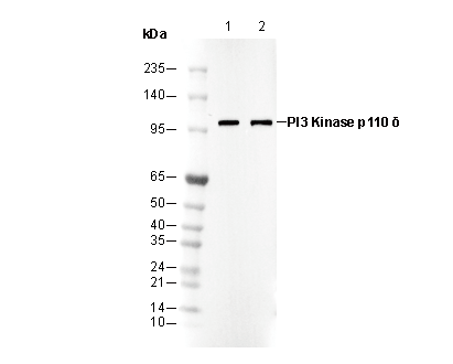 Lane 1: SW620
Lane 2: HT1080