|
Wie zu zitieren 1. Für Zitate im Text (Materialien & Methoden): 2. Für die Tabelle der Schlüsselressourcen: |
||
|
Gebührenfrei: (877) 796-6397 -- Nur USA und Kanada -- |
Fax: +1-832-582-8590 Bestellungen: +1-832-582-8158 |
Technischer Support: +1-832-582-8158 Ext:3 Bitte geben Sie Ihre Bestellnummer in der E-Mail an. Wir bemühen uns, alle E-Mail-Anfragen innerhalb eines Werktages zu beantworten. |
Biologische Beschreibung
| Spezifität | RNF40 Antibody [D22C8] erkennt endogene Spiegel des gesamten RNF40-Proteins. Dieser Antikörper kreuzreagiert nicht mit RNF20/BRE1A. |
|---|---|
| Hintergrund | RNF40 ist eine RING-Finger-E3 Ubiquitin Ligase mit mehreren Coiled-Coil-Domänen und einem C-terminalen RING-Finger-Motiv, das an Protein-DNA- und Protein-Protein-Interaktionen beteiligt ist. Es kodiert ein 1001 Aminosäuren umfassendes Polypeptid mit einer Masse von 113.678 Da. RNF40 bildet einen stabilen Heterodimer mit seinem Paralog RNF20, das Histon H2B an Lysin 120 und andere Nicht-Histon-Proteine monoubiquityliert. RNF40 fungiert sowohl als Tumorsuppressor als auch als Onkogen und spielt eine entscheidende Rolle bei der Krebsentwicklung, -progression und -metastasierung. RNF40 befindet sich auf Chromosom 16p11.2 und wird ubiquitär in menschlichen und Ratten-Geweben exprimiert. Es ist essenziell für die Monoubiquitinierung von Histon 2B an Lysin 120 (H2Bub1) und beeinflusst Stammzelleneigenschaften in normalem Brustgewebe und Basal-ähnlichem Brustkrebs (BLBC). RNF40 hat anti-apoptotische und pro-inflammatorische Rollen bei Darmkrebs und unterstützt Tumoren, indem es die Apoptose unterdrückt und die Aktivität der fokalen Adhäsionskinase (FAK) bei HER2+-Brustkrebs fördert. |
Nutzungsinformationen
| Anwendung | WB, IP | Verdünnung |
|
||||
|---|---|---|---|---|---|---|---|
| Reaktivität | Human | ||||||
| Quelle | Rabbit Monoclonal Antibody | MW | 130 kDa | ||||
| Lagerpuffer | PBS, pH 7.2+50% Glycerol+0.05% BSA+0.01% NaN₃ | Lagerung (Ab dem Datum des Erhalts) |
–20°C (avoid freeze-thaw cycles), 2 years | ||||
| WB |
Experimental Protocol:
Sample preparation
1. Tissue: Lyse the tissue sample by adding an appropriate volume of ice-cold RIPA/Nuclear Lysis Buffer (containing Protease Inhibitor Cocktail),and homogenize the tissue at a low temperature or lyse it by sonication on ice, then incubate on ice for 30 minutes. 2. Adherent cell: Aspirate the culture medium and transfer the cells into an EP tube. Wash the cells with ice-cold PBS twice. Add an appropriate volume of RIPA/Nuclear Lysis Buffer (containing Protease Inhibitor Cocktail), sonicate to lyse the cells, and incubate on ice for 30 minutes. 3. Suspension cell: Transfer the culture medium to a pre-cooled centrifuge tube. Centrifuge and aspirate the supernatant. Wash the cells with ice-cold PBS twice.Add an appropriate volume of RIPA/Nuclear Lysis Buffer (containing Protease Inhibitor Cocktail), sonicate to lyse the cells, and incubate on ice for 30 minutes. 4. Place the lysate into a pre-cooled microcentrifuge tube. Centrifuge at 4°C for 15 min. Collect the supernatant;
5. Remove a small volume of lysate to determine the protein concentration;
6. Combine the lysate with protein loading buffer. Boil 20 µL sample under 95-100°C for 5 min. Centrifuge for 5 min after cool down on ice.
Electrophoretic separation
1. According to the concentration of extracted protein, load appropriate amount of protein sample and marker onto SDS-PAGE gels for electrophoresis. Recommended separating gel (lower gel) concentration: 5%. Reference Table for Selecting SDS-PAGE Separation Gel Concentrations 2. Power up 80V for 30 minutes. Then the power supply is adjusted (110 V~150 V), the Marker is observed, and the electrophoresis can be stopped when the indicator band of the predyed protein Marker where the protein is located is properly separated. (Note that the current should not be too large when electrophoresis, too large current (more than 150 mA) will cause the temperature to rise, affecting the result of running glue. If high currents cannot be avoided, an ice bath can be used to cool the bath.)
Transfer membrane
1. Take out the converter, soak the clip and consumables in the pre-cooled converter;
2. Activate PVDF membrane with methanol for 1 min and rinse with transfer buffer;
3. Install it in the order of "black edge of clip - sponge - filter paper - filter paper - glue -PVDF membrane - filter paper - filter paper - sponge - white edge of clip"; 4. The protein was electrotransferred to PVDF membrane. ( 0.45 µm PVDF membrane is recommended ) Reference Table for Selecting PVDF Membrane Pore Size Specifications Recommended conditions for wet transfer: 200 mA, 120 min. ( Note that the transfer conditions can be adjusted according to the protein size. For high-molecular-weight proteins, a higher current and longer transfer time are recommended. However, ensure that the transfer tank remains at a low temperature to prevent gel melting.)
Block
1. After electrotransfer, wash the film with TBST at room temperature for 5 minutes;
2. Incubate the film in the blocking solution for 1 hour at room temperature;
3. Wash the film with TBST for 3 times, 5 minutes each time.
Antibody incubation
1. Use 5% skim milk powder to prepare the primary antibody working liquid (recommended dilution ratio for primary antibody 1:1000), gently shake and incubate with the film at 4°C overnight; 2. Wash the film with TBST 3 times, 5 minutes each time;
3. Add the secondary antibody to the blocking solution and incubate with the film gently at room temperature for 1 hour;
4. After incubation, wash the film with TBST 3 times for 5 minutes each time.
Antibody staining
652. Add the prepared ECL luminescent substrate (or select other color developing substrate according to the second antibody) and mix evenly;
2. Incubate with the film for 1 minute, remove excess substrate (keep the film moist), wrap with plastic film, and expose in the imaging system.
|
Referenzen
|
Anwendungsdaten
WB
Validiert von Selleck
-
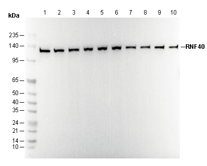 Lane 1: HCT 116
Lane 2: DLD-1
Lane 3: MCF7
Lane 4: OVCAR8
Lane 5: A172
Lane 6: Hela
Lane 7: DU145
Lane 8: HPB-ALL
Lane 9: A-375
Lane 10: RH30