|
Wie zu zitieren 1. Für Zitate im Text (Materialien & Methoden): 2. Für die Tabelle der Schlüsselressourcen: |
||
|
Gebührenfrei: (877) 796-6397 -- Nur USA und Kanada -- |
Fax: +1-832-582-8590 Bestellungen: +1-832-582-8158 |
Technischer Support: +1-832-582-8158 Ext:3 Bitte geben Sie Ihre Bestellnummer in der E-Mail an. Wir bemühen uns, alle E-Mail-Anfragen innerhalb eines Werktages zu beantworten. |
Biologische Beschreibung
| Spezifität | TCF12/HEB Antibody [G2F21] detektiert endogene Spiegel des gesamten TCF12/HEB-Proteins. |
|---|---|
| Hintergrund | TCF12/HEB (Transkriptionsfaktor 12/HeLa E-box Binding protein) ist ein Klasse-I-E-Protein-Transkriptionsfaktor mit basischer Helix-Loop-Helix-Struktur (bHLH), der von TCF12 auf Chromosom 15q21 kodiert wird und in zwei Hauptisoformen existiert: HEBCan (681 Aminosäuren, vom ubiquitären Promotor) und HEBAlt (kurze Form, von einem distalen Promotor). Diese Isoformen heterodimerisieren mit gewebespezifischen bHLH-Partnern wie E47, MyoD und NeuroD über ihre konservierte bHLH-Domäne, die aus einer basischen DNA-Bindungsregion und einem HLH-Motiv für die Dimerisierung besteht, um CANNTG E-Box-Konsensussequenzen zu erkennen und so die Abstammungsfestlegung in T-/B-Zellen, Muskeln und Neuronen zu steuern. TCF12/HEB enthält eine N-terminale Aktivierungsdomäne, die reich an sauren Resten für die Rekrutierung von Coaktivatoren ist, eine zentrale Transaktivierungsdomäne mit Glutamin-/Prolin-Strecken, die das Pausieren und die Freisetzung der RNA-Polymerase II verstärken, die zentrale bHLH-Domäne (~60 Reste), bei der die basische Helix die große Furche der DNA kontaktiert, während HLH-amphipathische Helices durch hydrophobe Grenzflächen dimerisieren, und C-terminale Hemmdomänen, die die Partnerspezifität modulieren. Alternatives Spleißen an Exon 1 erzeugt HEBCan für breite T-Zell-Entwicklungsstadien und HEBAlt, das in DN2/DN3-Thymocyten angereichert ist, um eine effiziente Vorläuferzellengeneration zu fördern. TCF12/HEB orchestriert die T-Zell-Entwicklung, indem es Lmo2 und Lyl1 an Eβ- und Cd4-Enhancern kofindet, um Gene zu aktivieren, die an Rekombination und Expansion beteiligt sind, reprimiert E2A-Ziele durch kompetitive Dimerisierung, um die Selbsterneuerung zu begrenzen, und gleicht die hämatopoetische Stammzell-(HSC)-Rekonstitution versus Differenzierung aus, wobei ein Mangel zu myeloischer Verzerrung, B-/T-Zell-Blockade und Proliferationsdefekten führt. Die PKCθ/Carma1-Signalgebung phosphoryliert HEBCan an Serinresten, wodurch die Id-Protein-vermittelte Autoinhibition aufgehoben und die TCR-induzierte Chromatin-Looping mit Runx1 und Foxp1 für die Il2ra- und Dtx1-Expression ermöglicht wird, während HEBAlt einzigartig mit Bcl11b am Tcrα-Enhancer zusammenarbeitet, um die positive Selektion zu erleichtern. TCF12-Mutationen verursachen koronale Kraniosynostose (Saethre-Chotzen-Syndrom) durch Störung der Twist1-Heterodimerisierung und der Suturenpatenz; Haploinsuffizienz ist durch neuroentwicklungsbedingte Gen-Dysregulation mit Dyslexie verbunden, und somatische Veränderungen tragen zur Leukämie (AML1-ETO-Fusionen) oder zur Progression solider Tumoren über EMT und Invasion bei. |
Nutzungsinformationen
| Anwendung | WB, IP, IHC | Verdünnung |
|
||||||
|---|---|---|---|---|---|---|---|---|---|
| Reaktivität | Human | ||||||||
| Quelle | Rabbit Monoclonal Antibody | MW | 85 kDa | ||||||
| Lagerpuffer | PBS, pH 7.2+50% Glycerol+0.05% BSA+0.01% NaN3 | Lagerung (Ab dem Datum des Erhalts) |
-20°C (avoid freeze-thaw cycles), 2 years | ||||||
| WB |
Experimental Protocol:
Sample preparation
1. Tissue: Lyse the tissue sample by adding an appropriate volume of ice-cold RIPA/Nuclear Lysis Buffer (containing Protease Inhibitor Cocktail),and homogenize the tissue at a low temperature. 2. Adherent cell: Aspirate the culture medium and wash the cells with ice-cold PBS twice. Lyse the cells by adding an appropriate volume of RIPA/Nuclear Lysis Buffer (containing Protease Inhibitor Cocktail) and put the sample on ice for 5 min. 3. Suspension cell: Transfer the culture medium to a pre-cooled centrifuge tube. Centrifuge and aspirate the supernatant. Wash the cells with ice-cold PBS twice. Lyse the cells by adding an appropriate volume of RIPA/Nuclear Lysis Buffer (containing Protease Inhibitor Cocktail) and put the sample on ice for 5 min. 4. Place the lysate into a pre-cooled microcentrifuge tube. Centrifuge at 4°C for 15 min. Collect the supernatant;
5. Remove a small volume of lysate to determine the protein concentration;
6. Combine the lysate with protein loading buffer. Boil 20 µL sample under 95-100°C for 5 min. Centrifuge for 5 min after cool down on ice.
Electrophoretic separation
1. According to the concentration of extracted protein, load appropriate amount of protein sample and marker onto SDS-PAGE gels for electrophoresis. Recommended separating gel (lower gel) concentration: 10%. Reference Table for Selecting SDS-PAGE Separation Gel Concentrations 2. Power up 80V for 30 minutes. Then the power supply is adjusted (110 V~150 V), the Marker is observed, and the electrophoresis can be stopped when the indicator band of the predyed protein Marker where the protein is located is properly separated. (Note that the current should not be too large when electrophoresis, too large current (more than 150 mA) will cause the temperature to rise, affecting the result of running glue. If high currents cannot be avoided, an ice bath can be used to cool the bath.)
Transfer membrane
1. Take out the converter, soak the clip and consumables in the pre-cooled converter;
2. Activate PVDF membrane with methanol for 1 min and rinse with transfer buffer;
3. Install it in the order of "black edge of clip - sponge - filter paper - filter paper - glue -PVDF membrane - filter paper - filter paper - sponge - white edge of clip"; 4. The protein was electrotransferred to PVDF membrane. ( 0.45 µm PVDF membrane is recommended ) Reference Table for Selecting PVDF Membrane Pore Size Specifications Recommended conditions for wet transfer: 200 mA, 120 min. ( Note that the transfer conditions can be adjusted according to the protein size. For high-molecular-weight proteins, a higher current and longer transfer time are recommended. However, ensure that the transfer tank remains at a low temperature to prevent gel melting.)
Block
1. After electrotransfer, wash the film with TBST at room temperature for 5 minutes;
2. Incubate the film in the blocking solution for 1 hour at room temperature;
3. Wash the film with TBST for 3 times, 5 minutes each time.
Antibody incubation
1. Use 5% skim milk powder to prepare the primary antibody working liquid (recommended dilution ratio for primary antibody 1:1000), gently shake and incubate with the film at 4°C overnight; 2. Wash the film with TBST 3 times, 5 minutes each time;
3. Add the secondary antibody to the blocking solution and incubate with the film gently at room temperature for 1 hour;
4. After incubation, wash the film with TBST 3 times for 5 minutes each time.
Antibody staining
1. Add the prepared ECL luminescent substrate (or select other color developing substrate according to the second antibody) and mix evenly;
2. Incubate with the film for 1 minute, remove excess substrate (keep the film moist), wrap with plastic film, and expose in the imaging system.
|
| IHC |
Experimental Protocol:
Deparaffinization/Rehydration
1. Deparaffinize/hydrate sections:
2. Incubate sections in three washes of xylene for 5 min each.
3. Incubate sections in two washes of 100% ethanol for 10 min each.
4. Incubate sections in two washes of 95% ethanol for 10 min each.
5. Wash sections two times in dH2O for 5 min each.
6.Antigen retrieval: For Citrate: Heat slides in a microwave submersed in 1X citrate unmasking solution until boiling is initiated; continue with 10 min at a sub-boiling temperature (95°-98°C). Cool slides on bench top for 30 min.
Staining
1. Wash sections in dH2O three times for 5 min each.
2. Incubate sections in 3% hydrogen peroxide for 10 min.
3. Wash sections in dH2O two times for 5 min each.
4. Wash sections in wash buffer for 5 min.
5. Block each section with 100–400 µl of blocking solution for 1 hr at room temperature.
6. Remove blocking solution and add 100–400 µl primary antibody diluent in to each section. Incubate overnight at 4°C.
7. Remove antibody solution and wash sections with wash buffer three times for 5 min each.
8. Cover section with 1–3 drops HRPas needed. Incubate in a humidified chamber for 30 min at room temperature.
9. Wash sections three times with wash buffer for 5 min each.
10. Add DAB Chromogen Concentrate to DAB Diluent and mix well before use.
11. Apply 100–400 µl DAB to each section and monitor closely. 1–10 min generally provides an acceptable staining intensity.
12. Immerse slides in dH2O.
13. If desired, counterstain sections with hematoxylin.
14. Wash sections in dH2O two times for 5 min each.
15. Dehydrate sections: Incubate sections in 95% ethanol two times for 10 sec each; Repeat in 100% ethanol, incubating sections two times for 10 sec each; Repeat in xylene, incubating sections two times for 10 sec each.
16. Mount sections with coverslips and mounting medium.
|
Referenzen
|
Anwendungsdaten
WB
Validiert von Selleck
-
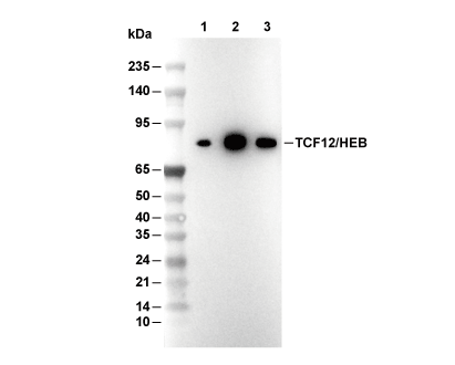 Lane 1: IMR-32, Lane 2: Jurkat, Lane 3: MOLT4