|
Wie zu zitieren 1. Für Zitate im Text (Materialien & Methoden): 2. Für die Tabelle der Schlüsselressourcen: |
||
|
Gebührenfrei: (877) 796-6397 -- Nur USA und Kanada -- |
Fax: +1-832-582-8590 Bestellungen: +1-832-582-8158 |
Technischer Support: +1-832-582-8158 Ext:3 Bitte geben Sie Ihre Bestellnummer in der E-Mail an. Wir bemühen uns, alle E-Mail-Anfragen innerhalb eines Werktages zu beantworten. |
Biologische Beschreibung
| Spezifität | TIMP3 Antibody [C11F19] weist endogene Spiegel des gesamten TIMP3-Proteins nach. |
|---|---|
| Hintergrund | TIMP3 (Tissue Inhibitor of Metalloproteinases 3) ist ein einzigartiges Mitglied der TIMP-Familie, das durch das TIMP3-Gen kodiert wird und sich durch seine starke Bindungsaffinität zur extrazellulären Matrix (ECM) und seine breite Hemmwirkung gegen Matrix-Metalloproteinasen (MMPs), ADAMs und ADAMTSs auszeichnet. Es spielt eine entscheidende Rolle bei der Aufrechterhaltung der ECM-Homöostase und der Regulierung der Geweberemodellierung. Das TIMP3-Protein besteht aus einer N-terminalen Domäne (~120 Aminosäuren) und einer C-terminalen Domäne, die durch sechs Disulfidbindungen zwischen zwölf konservierten Cysteinresten stabilisiert wird; es besitzt eine keilförmige Gestalt, die in die aktiven Zentren von Metalloproteinasen passt, um deren proteolytische Aktivitäten zu hemmen. TIMP3 hemmt die Angiogenese, reguliert die Apoptose und erhält die ECM-Integrität, indem es an Matrixkomponenten bindet und so Zell-Matrix-Interaktionen moduliert. Es ist an Signalwegen wie NF-κB beteiligt und gleicht Entzündungen und Geweberemodellierung durch die Regulierung wichtiger Proteine wie TNF-alpha und Metalloproteinasen aus. Eine Dysregulation von TIMP3 ist an verschiedenen Krankheiten beteiligt, darunter chronisch-obstruktive Lungenerkrankung (COPD), altersbedingte Makuladegeneration (AMD), Sorsby-Fundusdystrophie, diabetische Nephropathie und Osteoarthritis, oft durch gestörten ECM-Umsatz und aberrante Angiogenese. |
Nutzungsinformationen
| Anwendung | WB | Verdünnung |
|
||
|---|---|---|---|---|---|
| Reaktivität | Human, Mouse, Rat | ||||
| Quelle | Rabbit Monoclonal Antibody | MW | 20 kDa, 25 kDa | ||
| Lagerpuffer | PBS, pH 7.2+50% Glycerol+0.05% BSA+0.01% NaN3 | Lagerung (Ab dem Datum des Erhalts) |
-20°C (avoid freeze-thaw cycles), 2 years | ||
| WB |
Experimental Protocol:
Sample preparation
1. Tissue: Lyse the tissue sample by adding an appropriate volume of ice-cold RIPA/NP-40 Lysis Buffer (containing Protease Inhibitor Cocktail),and homogenize the tissue at a low temperature. 2. Adherent cell: Aspirate the culture medium and wash the cells with ice-cold PBS twice. Lyse the cells by adding an appropriate volume of RIPA/NP-40 Lysis Buffer (containing Protease Inhibitor Cocktail) and put the sample on ice for 5 min. 3. Suspension cell: Transfer the culture medium to a pre-cooled centrifuge tube. Centrifuge and aspirate the supernatant. Wash the cells with ice-cold PBS twice. Lyse the cells by adding an appropriate volume of RIPA/NP-40 Lysis Buffer (containing Protease Inhibitor Cocktail) and put the sample on ice for 5 min. 4. Place the lysate into a pre-cooled microcentrifuge tube. Centrifuge at 4°C for 15 min. Collect the supernatant;
5. Remove a small volume of lysate to determine the protein concentration;
6. Combine the lysate with protein loading buffer. Boil 20 µL sample under 95-100°C for 5 min. Centrifuge for 5 min after cool down on ice.
Electrophoretic separation
1. According to the concentration of extracted protein, load appropriate amount of protein sample and marker onto SDS-PAGE gels for electrophoresis. Recommended separating gel (lower gel) concentration: 10%. Reference Table for Selecting SDS-PAGE Separation Gel Concentrations 2. Power up 80V for 30 minutes. Then the power supply is adjusted (110 V~150 V), the Marker is observed, and the electrophoresis can be stopped when the indicator band of the predyed protein Marker where the protein is located is properly separated. (Note that the current should not be too large when electrophoresis, too large current (more than 150 mA) will cause the temperature to rise, affecting the result of running glue. If high currents cannot be avoided, an ice bath can be used to cool the bath.)
Transfer membrane
1. Take out the converter, soak the clip and consumables in the pre-cooled converter;
2. Activate PVDF membrane with methanol for 1 min and rinse with transfer buffer;
3. Install it in the order of "black edge of clip - sponge - filter paper - filter paper - glue -PVDF membrane - filter paper - filter paper - sponge - white edge of clip"; 4. The protein was electrotransferred to PVDF membrane. ( 0.45 µm PVDF membrane is recommended ) Reference Table for Selecting PVDF Membrane Pore Size Specifications Recommended conditions for wet transfer: 200 mA, 60 min. ( Note that the transfer conditions can be adjusted according to the protein size. For high-molecular-weight proteins, a higher current and longer transfer time are recommended. However, ensure that the transfer tank remains at a low temperature to prevent gel melting.)
Block
1. After electrotransfer, wash the film with TBST at room temperature for 5 minutes;
2. Incubate the film in the blocking solution for 1 hour at room temperature;
3. Wash the film with TBST for 3 times, 5 minutes each time.
Antibody incubation
1. Use 5% skim milk powder to prepare the primary antibody working liquid (recommended dilution ratio for primary antibody 1:1000), gently shake and incubate with the film at 4°C overnight; 2. Wash the film with TBST 3 times, 5 minutes each time;
3. Add the secondary antibody to the blocking solution and incubate with the film gently at room temperature for 1 hour;
4. After incubation, wash the film with TBST 3 times for 5 minutes each time.
Antibody staining
1. Add the prepared ECL luminescent substrate (or select other color developing substrate according to the second antibody) and mix evenly;
2. Incubate with the film for 1 minute, remove excess substrate (keep the film moist), wrap with plastic film, and expose in the imaging system.
|
Referenzen
|
Anwendungsdaten
WB
Validiert von Selleck
-
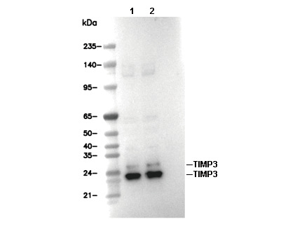 Lane 1: 3T3, Lane 2: A431