|
Wie zu zitieren 1. Für Zitate im Text (Materialien & Methoden): 2. Für die Tabelle der Schlüsselressourcen: |
||
|
Gebührenfrei: (877) 796-6397 -- Nur USA und Kanada -- |
Fax: +1-832-582-8590 Bestellungen: +1-832-582-8158 |
Technischer Support: +1-832-582-8158 Ext:3 Bitte geben Sie Ihre Bestellnummer in der E-Mail an. Wir bemühen uns, alle E-Mail-Anfragen innerhalb eines Werktages zu beantworten. |
Biologische Beschreibung
| Spezifität | Tin2 Antibody [K6G20] weist endogene Spiegel des gesamten Tin2-Proteins nach. |
|---|---|
| Hintergrund | TIN2 (TRF1-interacting nuclear factor 2) ist ein zentrales Gerüstprotein innerhalb des Shelterin-Komplexes, einer Sechs-Protein-Anordnung, die die Telomere von Säugetieren schützt und die Telomerlänge und -integrität reguliert. TIN2 enthält eine Telomer-Repeat-Faktor-Homologie-(TRFH)-Domäne, ein TRFH-Bindungsmotiv (TBM) und eine Domäne, die mit Mutationen der Dyskeratosis congenita assoziiert ist. TIN2 interagiert über seine TBM- und TRFH-Domänen mit TRF1 und TRF2, doppelsträngigen telomeren DNA-Bindungsproteinen, und bindet direkt an TPP1, um die Ansammlung von POT1 und TPP1 an den Telomeren zu erleichtern, was für den Schutz der einzelsträngigen telomeren DNA entscheidend ist. Es gibt zwei Haupt-TIN2-Isoformen, TIN2S und TIN2L, die die Telomerstabilität und -architektur modulieren. TIN2 koordiniert Cis- (DNA-Kompaktierung) und Trans- (DNA-DNA- oder DNA-RNA-Brückenbildung) Interaktionen und erhält die strukturelle Integrität der Telomere aufrecht. Der Verlust von TIN2 führt zu Telomer-Deprotektion, Aktivierung von DNA Damage/DNA Repair-Antworten (ATM/ATR-Kinasen), Chromatidfusion und kann zu embryonaler Letalität führen. TIN2 interagiert auch mit der Cohesin-Untereinheit SA1 und reguliert die Schwestertelomer-Kohäsion, während Tankyrase1 die TRF1-TIN2-Assoziation mit Telomeren kontrolliert. Als kooperative Plattform überbrückt TIN2 TRF1, TRF2 und TPP1 und ermöglicht so die Shelterin-Funktionen beim Chromosomenendschutz, der Telomerase-Regulation und der Genomstabilität. Mutationen oder Fehlfunktionen von TIN2 sind mit vorzeitigen Alterungssyndromen und Krebsanfälligkeit verbunden. |
Nutzungsinformationen
| Anwendung | WB | Verdünnung |
|
||
|---|---|---|---|---|---|
| Reaktivität | Mouse, Rat, Human | ||||
| Quelle | Rabbit Monoclonal Antibody | MW | 50 kDa | ||
| Lagerpuffer | PBS, pH 7.2+50% Glycerol+0.05% BSA+0.01% NaN3 | Lagerung (Ab dem Datum des Erhalts) |
-20°C (avoid freeze-thaw cycles), 2 years | ||
| WB |
Experimental Protocol:
Sample preparation
1. Tissue: Lyse the tissue sample by adding an appropriate volume of ice-cold RIPA/Nuclear Lysis Buffer (containing Protease Inhibitor Cocktail),and homogenize the tissue at a low temperature. 2. Adherent cell: Aspirate the culture medium and wash the cells with ice-cold PBS twice. Lyse the cells by adding an appropriate volume of RIPA/Nuclear Lysis Buffer (containing Protease Inhibitor Cocktail) and put the sample on ice for 5 min. 3. Suspension cell: Transfer the culture medium to a pre-cooled centrifuge tube. Centrifuge and aspirate the supernatant. Wash the cells with ice-cold PBS twice. Lyse the cells by adding an appropriate volume of RIPA/Nuclear Lysis Buffer (containing Protease Inhibitor Cocktail) and put the sample on ice for 5 min. 4. Place the lysate into a pre-cooled microcentrifuge tube. Centrifuge at 4°C for 15 min. Collect the supernatant;
5. Remove a small volume of lysate to determine the protein concentration;
6. Combine the lysate with protein loading buffer. Boil 20 µL sample under 95-100°C for 5 min. Centrifuge for 5 min after cool down on ice.
Electrophoretic separation
1. According to the concentration of extracted protein, load appropriate amount of protein sample and marker onto SDS-PAGE gels for electrophoresis. Recommended separating gel (lower gel) concentration: 10%. Reference Table for Selecting SDS-PAGE Separation Gel Concentrations 2. Power up 80V for 30 minutes. Then the power supply is adjusted (110 V~150 V), the Marker is observed, and the electrophoresis can be stopped when the indicator band of the predyed protein Marker where the protein is located is properly separated. (Note that the current should not be too large when electrophoresis, too large current (more than 150 mA) will cause the temperature to rise, affecting the result of running glue. If high currents cannot be avoided, an ice bath can be used to cool the bath.)
Transfer membrane
1. Take out the converter, soak the clip and consumables in the pre-cooled converter;
2. Activate PVDF membrane with methanol for 1 min and rinse with transfer buffer;
3. Install it in the order of "black edge of clip - sponge - filter paper - filter paper - glue -PVDF membrane - filter paper - filter paper - sponge - white edge of clip"; 4. The protein was electrotransferred to PVDF membrane. ( 0.45 µm PVDF membrane is recommended ) Reference Table for Selecting PVDF Membrane Pore Size Specifications Recommended conditions for wet transfer: 200 mA, 120 min. ( Note that the transfer conditions can be adjusted according to the protein size. For high-molecular-weight proteins, a higher current and longer transfer time are recommended. However, ensure that the transfer tank remains at a low temperature to prevent gel melting.)
Block
1. After electrotransfer, wash the film with TBST at room temperature for 5 minutes;
2. Incubate the film in the blocking solution for 1 hour at room temperature;
3. Wash the film with TBST for 3 times, 5 minutes each time.
Antibody incubation
1. Use 5% skim milk powder to prepare the primary antibody working liquid (recommended dilution ratio for primary antibody 1:1000), gently shake and incubate with the film at 4°C overnight; 2. Wash the film with TBST 3 times, 5 minutes each time;
3. Add the secondary antibody to the blocking solution and incubate with the film gently at room temperature for 1 hour;
4. After incubation, wash the film with TBST 3 times for 5 minutes each time.
Antibody staining
1. Add the prepared ECL luminescent substrate (or select other color developing substrate according to the second antibody) and mix evenly;
2. Incubate with the film for 1 minute, remove excess substrate (keep the film moist), wrap with plastic film, and expose in the imaging system.
|
Referenzen
|
Anwendungsdaten
WB
Validiert von Selleck
-
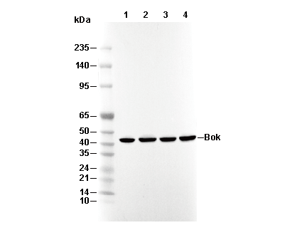 Lane 1: MCF7, Lane 2: C6, Lane 3: RAW264.7, Lane 4: PC12