Vadimezan (DMXAA)

Katalog-Nr.S1537 Charge:S153709

Drucken

Technische Daten

Formel

C17H14O4

Molekulargewicht 282.29 CAS-Nr. 117570-53-3
Löslichkeit (25°C)* In vitro DMSO 6 mg/mL (21.25 mM)
Water Insoluble
Ethanol Insoluble
In vivo (Lösungsmittel einzeln und der Reihe nach zum Produkt hinzufügen.)
Homogeneous suspension
CMC-NA
≥5mg/ml Taking the 1 mL working solution as an example, add 5 mg of this product to 1 ml of CMC-Na solution, mix evenly to obtain a homogeneous suspension with a final concentration of 5 mg/ml.
Clear solution
10% DMSO 40%PEG300 5%Tween80 45%ddH2O

Validiert von Selleck Labs. Sollten Sie Anpassungen an dieser Formulierung benötigen, wenden Sie sich an unser Vertriebsteam für kundenspezifische Tests.

0.750mg/ml (2.66mM) Taking the 1 mL working solution as an example, add 100 μL of 7.5 mg/ml clarified DMSO stock solution to 400 μL of PEG300, mix evenly to clarify it; add 50 μL of Tween80 to the above system, mix evenly to clarify; then continue to add 450 μL of ddH2O to adjust the volume to 1 mL. The mixed solution should be used immediately for optimal results. 
* <1 mg/ml bedeutet schwer löslich oder unlöslich.
* Bitte beachten Sie, dass Selleck die Löslichkeit aller Verbindungen intern testet und die tatsächliche Löslichkeit geringfügig von veröffentlichten Werten abweichen kann. Dies ist normal und ist auf geringfügige Batch-zu-Batch-Variationen zurückzuführen.
* Versand bei Raumtemperatur (Stabilitätstests zeigen, dass dieses Produkt ohne Kühlmaßnahmen versendet werden kann.)

Vorbereitung von Stammlösungen

Biologische Aktivität

Beschreibung Vadimezan (DMXAA) ist ein vascular disrupting agents (VDA) und ein kompetitiver Inhibitor der DT-Diaphorase mit einem Ki von 20 μM bzw. einer IC50 von 62,5 μM in zellfreien Assays. Es ist auch ein STING-Agonist mit potenzieller antineoplastischer Aktivität, der in vitro stark IFN-β, aber relativ wenig TNF-α-Expression induziert. Diese Verbindung hat Antiviral-Aktivität. Phase 3.
Ziele
DT-diaphorase
(Cell-free assay)
DT-diaphorase
(Cell-free assay)
20 μM(Ki) 20 μM(Ki)
In vitro In DLD-1-Zellen des menschlichen Kolonkarzinoms hemmt Vadimezan (DMXAA) die DT-Diaphorase-Aktivität ohne signifikante Auswirkungen auf die Aktivität der Cytochrom-b5-Reduktase und der Cytochrom-P450-Reduktase. Die Kombination von Menadion und dieser Verbindung führt zu einer Erhöhung der antiproliferativen Aktivität von DLD-1-Zellen. Als Antiviral-Mittel hemmt es die VSV-induzierte Zytotoxizität und die Influenza-Virusreplikation in RAW 264.7-Makrophagen. Eine aktuelle Studie zeigt, dass DMXAA nicht-immunvermittelte hemmende Effekte gegen mehrere Kinase-Mitglieder des VEGFR (vascular endothelial growth factor receptor) aufweist, wie z.B. die VEGFR2-Signalgebung in menschlichen Nabelschnurvenenendothelzellen.
In vivo Vadimezan (DMXAA)-Behandlung schützt C57BL/6J-Mäuse, die i.n. mit 200 p.f.u. Maus-adaptiertem H1N1-Influenza-PR8-Virus infiziert wurden, signifikant mit einer Überlebensrate von 60 %, während die Kontrollgruppe nur eine Überlebensrate von 20 % aufwies. Diese Verbindung verzögert signifikant das durch chemische Karzinogene induzierte Tumorwachstum, verlängert die Zeit bis zur Tumorverdopplung und verlängert die Zeit von der Behandlung bis zur Euthanasie. Nach ihrer Behandlung wurden die mediane Tumorverdopplungszeit, die mediane Tumorverdreifachungszeit und die mediane Zeit von der Behandlung bis zur Euthanasie bei tumortragenden Tieren um das ca. 4,4-, 1,8- bzw. 2,7-fache erhöht.

Protokoll (aus Referenz)

Kinase-Assay:[1]
  • DT-Diaphorase-Aktivität und kinetische Analyse der Enzyminhibition

    Die Aktivität des gereinigten DT-Diaphorase-Enzyms wird durch Messung der Reduktion von Cytochrom c bei 550 nm an einem Beckman DU 650 Spektrophotometer bestimmt. Jeder Assay enthält Cytochrom c (70 μM), NADH (variable Konzentrationen), gereinigte DT-Diaphorase (0,032 μg) und Menadion (variable Konzentrationen) in einem Endvolumen von 1 ml Tris-HCl-Puffer (50 mM, pH 7,4), der 0,14 % BSA enthält. Die Reaktion wird durch Zugabe von NADH gestartet. Die Reduktionsraten werden über den Anfangsteil der Reaktionskurve (30 Sekunden) berechnet, und die Ergebnisse werden in μmol Cytochrom c reduziert/min/mg Protein ausgedrückt, wobei ein molarer Extinktionskoeffizient von 21,1 mM−1 cm−1 für reduziertes Cytochrom c verwendet wird. Enzymtests werden bei Raumtemperatur durchgeführt und alle Reaktionen dreifach. Die Hemmung der Aktivität der gereinigten DT-Diaphorase erfolgt durch Zugabe von Vadimezan (DMXAA) in verschiedenen Konzentrationen zur Reaktion, und die Hemmungseigenschaften werden durch Variation der NADH-Konzentration (konstantes Menadion) oder der Menadion-Konzentration (konstantes NADH) bei mehreren Konzentrationen dieser Verbindung bestimmt. Ki-Werte werden durch Auftragen von 1/V gegen erhalten. Die Aktivität der DT-Diaphorase in DLD-1-Zellen wird durch Messung der Dicumarol-sensitiven Reduktion von DCPIP bei 600 nm bestimmt. Kurz gesagt, DLD-1-Zellen im mittleren exponentiellen Wachstum werden durch Abschaben in eiskalten Puffer (Tris-HCl, 25 mM, pH 7,4 und 250 mM Saccharose) geerntet und auf Eis sonifiziert. Die Enzymtestbedingungen sind 2 mM NADH, 40 μM DCPIP, 20 μl Dicumarol (falls erforderlich) in einem Endvolumen von 1 ml Tris-HCl (25 mM, pH 7,4), das BSA (0,7 mg/ml) enthält. Die Ergebnisse werden als Dicumarol-sensitive Reduktion von DCPIP unter Verwendung eines molaren Extinktionskoeffizienten von 21 mM−1 cm−1 ausgedrückt. Proteinkonzentrationen werden mit dem Bradford-Assay bestimmt.

Zell-Assay:[1]
  • Zelllinien

    DLD-1 and H460 cells

  • Konzentrationen

    0-2 mM

  • Inkubationszeit

    96 hours

  • Methode

    DLD-1 human colon carcinoma and H460 human non-small cell lung carcinoma cells are routinely maintained as monolayer cultures in RPMI 1640 culture medium supplemented with foetal calf serum (10%), sodium pyruvate (2 mM), penicillin/streptomycin (50 IU mL−1/50 μg mL-1) and l-glutamine (2 mM). Chemosensitivity is assessed using the MTT assay and all assays are performed under aerobic conditions. Briefly, cells are plated into each well of a 96-well culture plate and incubated overnight in an atmosphere containing 5% CO2. Culture medium is completely removed from each well and replaced with medium containing a range of drug concentrations. In the case of menadione alone, the duration of drug exposure is 1 hour, after which the cells are washed twice with Hanks' balanced salt solution prior to the addition of 200 μL fresh RPMI 1640 medium to each well of the plate. For Vadimezan (DMXAA) alone, the duration of exposure is 3 hours. Following a four-day incubation, cell survival is determined using the MTT assay. For combinations of this compound with menadione, cells are initially set up and a non-toxic (>95% cell survival) concentration of it is selected. Cells are then initially exposed to DMXAA (2 mM) for a period of 2 hours, following which the medium is removed and replaced with medium containing the inhibitor (at a constant concentration of 2 mM) and menadione (at a range of drug concentrations). Following a further 7-hour incubation, cells are washed twice with Hanks' balanced salt solution prior to the addition of growth medium.

Tierstudie:[4]
  • Tiermodelle

    Chemical carcinogen (NMU) is injected into female Wistar rats.

  • Dosierungen

    ≤300 mg/kg

  • Verabreichung

    Administered via i.p.

Referenzen

  • https://pubmed.ncbi.nlm.nih.gov/10423172/
  • https://pubmed.ncbi.nlm.nih.gov/21084628/
  • https://pubmed.ncbi.nlm.nih.gov/22142330/
  • https://pubmed.ncbi.nlm.nih.gov/16944150/

Kundenproduktvalidierung

(B and C) sh-scrambled or sh-ck2a–transducted L929 cells (B) and Raw cells (C) were stimulated by DMXAA (100 μg/ml) for various times. Cytosolic and nuclear extracts were prepared as described in Materials and Methods. Five percent of the cytosolic proteins and 20% of the nuclear proteins were resolved by 10% SDS-PAGE. Subsequently, immunoblotting was conducted by indicated Abs. The amounts of Tubulin and Lamin B1 in cytosol versus nuclei detected by respective Abs were used as internal control for fractionation.

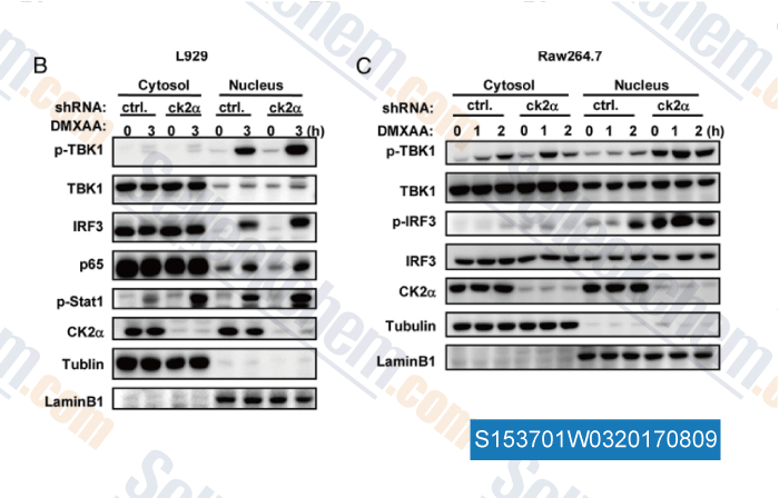
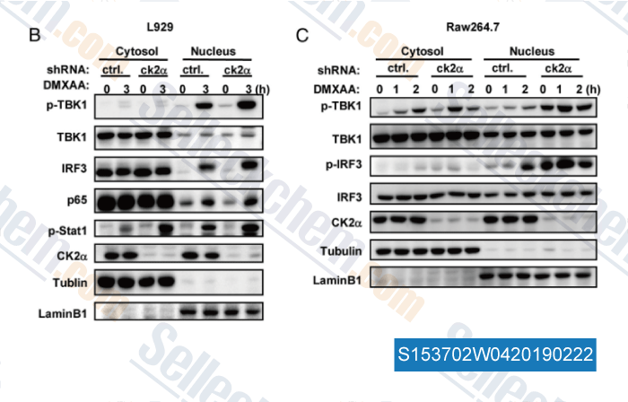
Daten von [ , , J Immunol, 2015, 194:4477-4488 ]

(E) WT and ASC−/− macrophages were stimulated with 1 μg/ml of tumor-derived DNA in the presence of lipofectamine or 50 μg/ml of DMXAA at different time points. Whole cell extracts were analysed with antibodies against pTBK1, total TBK1, pIRF3, total IRF3 and GAPDH.

Daten von [ , , J Immunol, 2016, 196(7):3191-8 ]

(B and C) sh-scrambled or sh-ck2α–transducted L929 cells (B) and Raw cells (C) were stimulated by DMXAA (100 μg/ml) for various times. Cytosolic and nuclear extracts were prepared as described in Materials and Methods. Five percent of the cytosolic proteins and 20% of the nuclear proteins were resolved by 10% SDS-PAGE. Subsequently, immunoblotting was conducted by indicated Abs. The amounts of Tubulin and Lamin B1 in cytosol versus nuclei detected by respective Abs were used as internal control for fractionation.

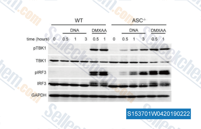
Daten von [ , , J Immunol, 2015, 194(9):4477-88 ]

(b-e) HEK293T cells were transiently transfected with empty vector (b), plasmids encoding hSTING(H232) (c), hSTING(R232) (d), or mSTING (e) together with IFNβ-luciferase reporter. After 24 h, cells were transfected with 2′,3′‐cGAMP (2 μg/mL) or stimulated with CMA (50 μg/mL), DMXAA (50 μM) and α-mangostin. Luciferase activity was measured 24 h after stimulation. Error bars represent the SD of independent experiments (n=3); *P<0.05, **P<0.01, ***P<0.001 (Student

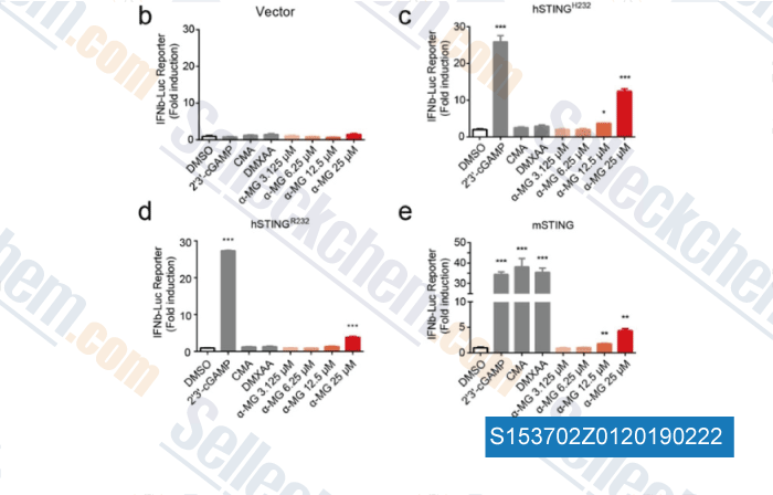
Daten von [ , , ChemMedChem, 2018, 13(19):2057-2064 ]

Sellecks Vadimezan (DMXAA) Wurde zitiert von 46 Publikationen

An oral tricyclic STING agonist suppresses tumor growth through remodeling of the immune microenvironment [ Cell Chem Biol, 2025, 32(2):280-290.e14] PubMed: 39904339
LAPTM5 exacerbates STING-mediated inflammation induced by LL-37 through stabilizing STING in rosacea [ Commun Biol, 2025, 8(1):1470] PubMed: 41087666
Profile of STING agonist and inhibitor research: a bibliometric analysis [ Front Pharmacol, 2025, 16:1528459] PubMed: 40008133
C9orf72 Alleviates DSS‑Induced Ulcerative Colitis via the cGAS-STING Pathway [ Immun Inflamm Dis, 2025, 13(1):e70139] PubMed: 39873292
YTHDF2 in peritumoral hepatocytes mediates chemotherapy-induced antitumor immune responses through CX3CL1-mediated CD8+ T cell recruitment [ Mol Cancer, 2024, 23(1):186] PubMed: 39237909
S-nitrosothiol homeostasis maintained by ADH5 facilitates STING-dependent host defense against pathogens [ Nat Commun, 2024, 15(1):1750] PubMed: 38409248
Mitochondrial DNA-boosted dendritic cell-based nanovaccination triggers antitumor immunity in lung and pancreatic cancers [ Cell Rep Med, 2024, 5(7):101648] PubMed: 38986624
FMT rescues mice from DSS-induced colitis in a STING-dependent manner [ Gut Microbes, 2024, 16(1):2397879] PubMed: 39324491
TBK1-Zyxin signaling controls tumor-associated macrophage recruitment to mitigate antitumor immunity [ EMBO J, 2024, 10.1038/s44318-024-00244-9] PubMed: 39304793
ISGylation by HERCs facilitates STING activation [ Cell Rep, 2024, 43(5):114135] PubMed: 38652662

RÜCKGABERICHTLINIE
Die bedingungslose Rückgaberichtlinie von Selleck Chemical gewährleistet unseren Kunden ein reibungsloses Online-Einkaufserlebnis. Wenn Sie in irgendeiner Weise mit Ihrem Kauf unzufrieden sind, können Sie jeden Artikel innerhalb von 7 Tagen nach Erhalt zurückgeben. Im Falle von Produktqualitätsproblemen, sei es protokollbezogene oder produktbezogene Probleme, können Sie jeden Artikel innerhalb von 365 Tagen ab dem ursprünglichen Kaufdatum zurückgeben. Bitte befolgen Sie die nachstehenden Anweisungen, wenn Sie Produkte zurücksenden.

VERSAND UND LAGERUNG
Selleck-Produkte werden bei Raumtemperatur transportiert. Wenn Sie das Produkt bei Raumtemperatur erhalten, seien Sie versichert, dass die Qualitätskontrollabteilung von Selleck Experimente durchgeführt hat, um zu überprüfen, dass die normale Temperaturplatzierung von einem Monat die biologische Aktivität von Pulverprodukten nicht beeinträchtigt. Nach dem Sammeln lagern Sie das Produkt bitte gemäß den in der Datenblatt beschriebenen Anforderungen. Die meisten Selleck-Produkte sind unter den empfohlenen Bedingungen stabil.

NICHT FÜR DIE ANWENDUNG AM MENSCHEN, FÜR VETERINÄRMEDIZINISCHE DIAGNOSTIK ODER THERAPEUTISCHE ZWECKE.