|
Wie zu zitieren 1. Für Zitate im Text (Materialien & Methoden): 2. Für die Tabelle der Schlüsselressourcen: |
||
|
Gebührenfrei: (877) 796-6397 -- Nur USA und Kanada -- |
Fax: +1-832-582-8590 Bestellungen: +1-832-582-8158 |
Technischer Support: +1-832-582-8158 Ext:3 Bitte geben Sie Ihre Bestellnummer in der E-Mail an. Wir bemühen uns, alle E-Mail-Anfragen innerhalb eines Werktages zu beantworten. |
Biologische Beschreibung
| Spezifität | VEGFB Antibody [E11E24] detektiert endogene Spiegel des gesamten VEGFB-Proteins. |
|---|---|
| Hintergrund | Vascular Endothelial Growth Factor B (VEGFB) ist ein homodimeres Zytokin, das zur PDGF/VEGF-Familie gehört und die Angiogenese fördert sowie den Lipidstoffwechsel in metabolisch aktiven Geweben wie Herz, Skelettmuskulatur und braunem Fettgewebe reguliert. VEGFB existiert in zwei Isoformen, VEGFB167 (heparinbindend, matrixassoziiert) und VEGFB186 (sezernierte Form), die beide ein konserviertes Cystinknotenmotiv mit sechs Cysteinresten aufweisen, die drei Disulfidbrücken bilden, welche die charakteristische VEGF-Dimerstruktur stabilisieren, die für die Rezeptorbindung essenziell ist. VEGFB bindet mit hoher Affinität an VEGFR-1 (Flt-1), hauptsächlich über spezifische Reste in seinen Rezeptorbindungsloops, wodurch Signalwege aktiviert werden, die das Überleben und die Proliferation von Endothelzellen unterstützen, ohne eine starke mitogene Aktivität im Vergleich zu VEGF-A aufzuweisen. VEGFB erhöht die Gefäßpermeabilität und -stabilisierung, während es den Fettsäuretransport über Endothelbarrieren einzigartig reguliert, indem es Translokaseproteine wie CD36 und FABP4/5 induziert und so die Blutgefäßbildung mit dem Gewebeenergiebedarf koordiniert. VEGFB signalisiert über VEGFR-1-Homodimere oder VEGFR-1/VEGFR-2-Heterodimere, aktiviert PI3K/Akt- und PLCγ-Signalwege, um die Gefäßreifung anstelle der sprießenden Angiogenese zu fördern. Dysreguliertes VEGFB trägt zu kardiovaskulären Pathologien bei, einschließlich ischämischer Herzkrankheit und Atherosklerose, und fungiert als therapeutisches Ziel zur Verbesserung der kardialen Vaskularisierung und der metabolischen Homöostase. |
Nutzungsinformationen
| Anwendung | WB | Verdünnung |
|
||
|---|---|---|---|---|---|
| Reaktivität | Rat, Mouse | ||||
| Quelle | Rabbit Monoclonal Antibody | MW | 22 kDa | ||
| Lagerpuffer | PBS, pH 7.2+50% Glycerol+0.05% BSA+0.01% NaN3 | Lagerung (Ab dem Datum des Erhalts) |
-20°C (avoid freeze-thaw cycles), 2 years | ||
| WB |
Experimental Protocol:
Sample preparation
1. Tissue: Lyse the tissue sample by adding an appropriate volume of ice-cold RIPA/NP-40 Lysis Buffer (containing Protease Inhibitor Cocktail),and homogenize the tissue at a low temperature. 2. Adherent cell: Aspirate the culture medium and wash the cells with ice-cold PBS twice. Lyse the cells by adding an appropriate volume of RIPA/NP-40 Lysis Buffer (containing Protease Inhibitor Cocktail) and put the sample on ice for 5 min. 3. Suspension cell: Transfer the culture medium to a pre-cooled centrifuge tube. Centrifuge and aspirate the supernatant. Wash the cells with ice-cold PBS twice. Lyse the cells by adding an appropriate volume of RIPA/NP-40 Lysis Buffer (containing Protease Inhibitor Cocktail) and put the sample on ice for 5 min. 4. Place the lysate into a pre-cooled microcentrifuge tube. Centrifuge at 4°C for 15 min. Collect the supernatant;
5. Remove a small volume of lysate to determine the protein concentration;
6. Combine the lysate with protein loading buffer. Boil 20 µL sample under 95-100°C for 5 min. Centrifuge for 5 min after cool down on ice.
Electrophoretic separation
1. According to the concentration of extracted protein, load appropriate amount of protein sample and marker onto SDS-PAGE gels for electrophoresis. Recommended separating gel (lower gel) concentration: 10%. Reference Table for Selecting SDS-PAGE Separation Gel Concentrations 2. Power up 80V for 30 minutes. Then the power supply is adjusted (110 V~150 V), the Marker is observed, and the electrophoresis can be stopped when the indicator band of the predyed protein Marker where the protein is located is properly separated. (Note that the current should not be too large when electrophoresis, too large current (more than 150 mA) will cause the temperature to rise, affecting the result of running glue. If high currents cannot be avoided, an ice bath can be used to cool the bath.)
Transfer membrane
1. Take out the converter, soak the clip and consumables in the pre-cooled converter;
2. Activate PVDF membrane with methanol for 1 min and rinse with transfer buffer;
3. Install it in the order of "black edge of clip - sponge - filter paper - filter paper - glue -PVDF membrane - filter paper - filter paper - sponge - white edge of clip"; 4. The protein was electrotransferred to PVDF membrane. ( 0.45 µm PVDF membrane is recommended ) Reference Table for Selecting PVDF Membrane Pore Size Specifications Recommended conditions for wet transfer: 200 mA, 60 min. ( Note that the transfer conditions can be adjusted according to the protein size. For high-molecular-weight proteins, a higher current and longer transfer time are recommended. However, ensure that the transfer tank remains at a low temperature to prevent gel melting.)
Block
1. After electrotransfer, wash the film with TBST at room temperature for 5 minutes;
2. Incubate the film in the blocking solution for 1 hour at room temperature;
3. Wash the film with TBST for 3 times, 5 minutes each time.
Antibody incubation
1. Use 5% skim milk powder to prepare the primary antibody working liquid (recommended dilution ratio for primary antibody 1:1000), gently shake and incubate with the film at 4°C overnight; 2. Wash the film with TBST 3 times, 5 minutes each time;
3. Add the secondary antibody to the blocking solution and incubate with the film gently at room temperature for 1 hour;
4. After incubation, wash the film with TBST 3 times for 5 minutes each time.
Antibody staining
1. Add the prepared ECL luminescent substrate (or select other color developing substrate according to the second antibody) and mix evenly;
2. Incubate with the film for 1 minute, remove excess substrate (keep the film moist), wrap with plastic film, and expose in the imaging system.
|
Referenzen
|
Anwendungsdaten
WB
Validiert von Selleck
-
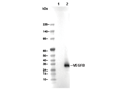 Lane 1: 293T, Lane 2: 293T (human VEGFB-3×FLAG transfected)