
nur für Forschungszwecke
Prostaglandin E2 (PGE2) Prostaglandin Receptor Agonist
Kat.-Nr.S3003
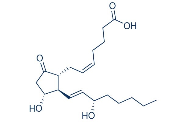
Chemische Struktur
Molekulargewicht: 352.47
Springe zu
Qualitätskontrolle
Charge:
Reinheit: >97%
- Zitiert in Nature Medicine für seine erstklassige Qualität
- COA
- Datenblatt
- SDS
- Zitiert in Nature Medicine für seine erstklassige Qualität
- COA
- Datenblatt
- SDS
- Zitiert in Nature Medicine für seine erstklassige Qualität
- COA
- NMR
- Datenblatt
- SDS
- Zitiert in Nature Medicine für seine erstklassige Qualität
- COA
- NMR
- Datenblatt
- SDS
- Zitiert in Nature Medicine für seine erstklassige Qualität
- COA
- NMR
- Datenblatt
- SDS
97
| Verwandte Ziele | Adrenergic Receptor Estrogen/progestogen Receptor GPR Androgen Receptor Glucocorticoid Receptor ACE RAAS Progesterone Receptor Opioid Receptor THR |
|---|---|
| Weitere PGES Inhibitoren | Yakuchinone A |
Zellkultur, Behandlung & Arbeitskonzentration
| Zelllinien | Assay-Typ | Konzentration | Inkubationszeit | Formulierung | Aktivitätsbeschreibung | PMID |
|---|---|---|---|---|---|---|
| HeLa | Function assay | TP_TRANSPORTER: uptake in PGT-expressing HeLa cells, Km = 0.094 μM. | 7754369 | |||
| HeLa | Function assay | TP_TRANSPORTER: uptake in PGT-expressing HeLa cells, K1/2 = 0.1 μM. | 8787677 | |||
| S2 | Function assay | TP_TRANSPORTER: uptake in OCT2-expressing S2 cells, Km = 0.0289 μM. | 11907186 | |||
| S2 | Function assay | TP_TRANSPORTER: uptake in OAT4-expressing S2 cells, Km = 0.154 μM. | 11907186 | |||
| S2 | Function assay | TP_TRANSPORTER: uptake in OAT3-expressing S2 cells, Km = 0.345 μM. | 11907186 | |||
| S2 | Function assay | TP_TRANSPORTER: uptake in OCT1-expressing S2 cells, Km = 0.657 μM. | 11907186 | |||
| S2 | Function assay | TP_TRANSPORTER: uptake in OAT2-expressing S2 cells, Km = 0.713 μM. | 11907186 | |||
| S2 | Function assay | TP_TRANSPORTER: uptake in OAT1-expressing S2 cells, Km = 0.97 μM. | 11907186 | |||
| HEK293 | Function assay | Inhibitory activity against human EP4 receptor expressed in HEK293 ebna cells, IC50 = 0.0007 μM. | 12643927 | |||
| HEK293 | Function assay | EP4 agonist potency utilizing a stable clone of pSV40-EP4 transfected into HEK293 cells expressing EP4 receptor, EC50 = 0.003 μM. | 12643927 | |||
| Sf9 | Function assay | TP_TRANSPORTER: uptake (vesicle) in membrane vesicles from MRP4-expressing Sf9 cells, Km = 3.4 μM. | 12835412 | |||
| HEK293 | Function assay | Displacement of [3H]PGE2 from human EP3 receptor expressed in HEK293 cells, Ki = 0.00033 μM. | 17531488 | |||
| HEK293 | Function assay | Displacement of [3H]PGE2 from human EP4 receptor expressed in HEK293 cells, Ki = 0.00079 μM. | 17531488 | |||
| HEK293 | Function assay | Displacement of [3H]PGE2 from human EP2 receptor expressed in HEK293 cells, Ki = 0.0049 μM. | 17531488 | |||
| HEK293 | Function assay | Displacement of [3H]PGE2 from human EP1 receptor expressed in HEK293 cells, Ki = 0.0091 μM. | 17531488 | |||
| HEK293 | Function assay | Agonist activity against rat EP2 receptor expressed in HEK293 cells assessed as stimulation of cAMP release, EC50 = 0.0002 μM. | 19250823 | |||
| HEK293 | Function assay | Agonist activity against rat EP4 receptor expressed in HEK293 cells assessed as stimulation of cAMP release, EC50 = 0.0007 μM. | 19250823 | |||
| HEK293 | Function assay | Inhibition of rat EP4 receptor expressed in HEK293 cells, IC50 = 0.0021 μM. | 19250823 | |||
| HEK293 | Function assay | Inhibition of rat EP2 receptor expressed in HEK293 cells, IC50 = 0.0052 μM. | 19250823 | |||
| CHO | Function assay | 60 mins | Displacement of [3H]-PGE2 from mouse EP4 receptor expressed in CHO cells after 60 mins by liquid scintillation counting, Ki = 0.0031 μM. | 22119471 | ||
| CHO | Function assay | 60 mins | Displacement of [3H]-PGE2 from mouse EP3 receptor expressed in CHO cells after 60 mins by liquid scintillation counting, Ki = 0.005 μM. | 22119471 | ||
| CHO | Function assay | 20 mins | Displacement of [3H]-PGE2 from mouse EP1 receptor expressed in CHO cells after 20 mins by liquid scintillation counting, Ki = 0.006 μM. | 22119471 | ||
| CHO | Function assay | 60 mins | Displacement of [3H]-PGE2 from mouse EP2 receptor expressed in CHO cells after 60 mins by liquid scintillation counting, Ki = 0.022 μM. | 22119471 | ||
| CHO | Function assay | 60 mins | Displacement of [3H]-PGE2 from mouse EP4 receptor expressed in CHO cells after 60 mins by scintillation counting, Ki = 0.0031 μM. | 22386979 | ||
| CHO | Function assay | 60 mins | Displacement of [3H]-PGE2 from mouse EP3 receptor expressed in CHO cells after 60 mins by scintillation counting, Ki = 0.005 μM. | 22386979 | ||
| CHO | Function assay | 60 mins | Displacement of [3H]-PGE2 from mouse EP1 receptor expressed in CHO cells after 60 mins by scintillation counting, Ki = 0.006 μM. | 22386979 | ||
| CHO | Function assay | 60 mins | Displacement of [3H]-PGE2 from mouse EP2 receptor expressed in CHO cells after 60 mins by scintillation counting, Ki = 0.022 μM. | 22386979 | ||
| CHEM1 | Function assay | 2 hrs | Displacement of [3H]PGE2 from human recombinant prostanoid EP4 receptor in CHEM1 cells after 2 hrs, Ki = 0.00045 μM. | 23403082 | ||
| CHEM1 | Function assay | 2 hrs | Displacement of [3H]PGE2 from human recombinant prostanoid EP4 receptor in CHEM1 cells after 2 hrs, IC50 = 0.0011 μM. | 23403082 | ||
| CHO | Function assay | 100 nM | Activity at recombinant EP1 receptor (unknown origin) expressed in CHO cells co-expressing Gq protein at 100 nM by electrical cell substrate impedance sensing system | 25701254 | ||
| CHO | Function assay | 100 nM | Activity at recombinant EP4 receptor (unknown origin) expressed in CHO cells co-expressing Gs protein at 100 nM by electrical cell substrate impedance sensing system | 25701254 | ||
| CHO | Function assay | 100 nM | Activity at recombinant EP3 receptor (unknown origin) expressed in CHO cells co-expressing Gi protein at 100 nM by electrical cell substrate impedance sensing system | 25701254 | ||
| CHO | Function assay | 30 mins | Agonist activity at human EP2 receptor expressed in CHO cells assessed as increase in intracellular cAMP level after 30 mins by HTRF method, EC50 = 0.0019 μM. | 26985320 | ||
| CHO | Function assay | Agonist activity at human EP3 receptor expressed in CHO cells assessed as increase in intracellular calcium level by fluorescence based analysis, EC50 = 0.0025 μM. | 26985320 | |||
| CHO | Function assay | Agonist activity at human EP1 receptor expressed in CHO cells assessed as increase in intracellular calcium level by fluorescence based analysis, EC50 = 0.0037 μM. | 26985320 | |||
| CHO | Function assay | 30 mins | Agonist activity at human EP4 receptor expressed in CHO cells assessed as increase in intracellular cAMP level after 30 mins by HTRF method, EC50 = 0.0075 μM. | 26985320 | ||
| Chem1 | Function assay | Agonist activity at human FP receptor expressed in human Chem1 cells assessed as increase in intracellular calcium level by fluorescence based analysis, EC50 = 0.25 μM. | 26985320 | |||
| HEK293 | Function assay | 90 mins | Agonist activity at PK2-tagged human EP2 receptor expressed in HEK293 cells assessed as induction of EA-tagged beta-arrestin recruitment incubated for 90 mins by beta-galactosidase reporter gene assay, EC50 = 0.346 μM. | 26985320 | ||
| CHO | Function assay | 30 mins | Agonist activity at human IP receptor expressed in CHO cells assessed as increase in intracellular cAMP level after 30 mins by HTRF method, EC50 = 0.347 μM. | 26985320 | ||
| HEK293 | Function assay | Displacement of [3H]PGE2 from human recombinant prostanoid EP2 receptor expressed in HEK293 cells, IC50 = 0.0026 μM. | 26988801 | |||
| HEK293 | Function assay | 120 mins | Displacement of [3H]PGE2 from human recombinant EP4 receptor expressed in HEK293 cells measured after 120 mins by scintillation counting method, IC50 = 0.00055 μM. | 27876250 | ||
| HEK293 | Function assay | 120 mins | Displacement of [3H]PGE2 from human recombinant EP2 receptor expressed in HEK293 cells measured after 120 mins by scintillation counting method, IC50 = 0.0026 μM. | 27876250 | ||
| Klicken Sie hier, um weitere experimentelle Daten zu Zelllinien anzuzeigen | ||||||
Chemische Informationen, Lagerung & Stabilität
| Molekulargewicht | 352.47 | Formel | C20H32O5 |
Lagerung (Ab dem Eingangsdatum) | |
|---|---|---|---|---|---|
| CAS-Nr. | 363-24-6 | SDF herunterladen | Lagerung von Stammlösungen | Lösungen sind instabil. Frisch zubereiten oder kleine, vorverpackte Größen kaufen. Nach Erhalt umpacken. | |
| Synonyme | Dinoprostone | Smiles | CCCCCC(C=CC1C(CC(=O)C1CC=CCCCC(=O)O)O)O | ||
Löslichkeit
|
In vitro |
DMSO
: 70 mg/mL
(198.59 mM)
Ethanol : 70 mg/mL Water : 2.5 mg/mL |
Molaritätsrechner
Verdünnungsrechner
Molekulargewichtsrechner
|
In vivo |
|||||
In-vivo-Formulierungsrechner (Klare Lösung)
Schritt 1: Geben Sie die untenstehenden Informationen ein (Empfohlen: Ein zusätzliches Tier zur Berücksichtigung von Verlusten während des Experiments)
mg/kg
g
μL
Schritt 2: Geben Sie die In-vivo-Formulierung ein (Dies ist nur der Rechner, keine Formulierung. Bitte kontaktieren Sie uns zuerst, wenn es im Abschnitt "Löslichkeit" keine In-vivo-Formulierung gibt.)
% DMSO
%
% Tween 80
% ddH2O
%DMSO
%
Berechnungsergebnisse:
Arbeitskonzentration: mg/ml;
Methode zur Herstellung der DMSO-Stammlösung: mg Wirkstoff vorgelöst in μL DMSO ( Konzentration der Stammlösung mg/mL, Bitte kontaktieren Sie uns zuerst, wenn die Konzentration die DMSO-Löslichkeit der Wirkstoffcharge überschreitet. )
Methode zur Herstellung der In-vivo-Formulierung: Nehmen Sie μL DMSO Stammlösung, dann hinzufügenμL PEG300, mischen und klären, dann hinzufügenμL Tween 80, mischen und klären, dann hinzufügen μL ddH2O, mischen und klären.
Methode zur Herstellung der In-vivo-Formulierung: Nehmen Sie μL DMSO Stammlösung, dann hinzufügen μL Maisöl, mischen und klären.
Hinweis: 1. Bitte stellen Sie sicher, dass die Flüssigkeit klar ist, bevor Sie das nächste Lösungsmittel hinzufügen.
2. Achten Sie darauf, das/die Lösungsmittel der Reihe nach hinzuzufügen. Sie müssen sicherstellen, dass die bei der vorherigen Zugabe erhaltene Lösung eine klare Lösung ist, bevor Sie mit der Zugabe des nächsten Lösungsmittels fortfahren. Physikalische Methoden wie Vortex, Ultraschall oder ein heißes Wasserbad können zur Unterstützung des Lösens verwendet werden.
Wirkmechanismus
| In vitro |
PGE2 wirkt über EP4-Rezeptoren auf Osteoklasten während des Fortschreitens von OA und OA-bedingten Schmerzen. |
|---|---|
| In vivo |
PGE2 (0,3 μg/kg, i.p.) reduziert signifikant die Anzahl der peritonealen Makrophagen, die in Ratten eine Phagozytose von Methacrylat-Mikrokügelchen durchführen. |
Literatur |
Klinische Studieninformationen
(Daten von https://clinicaltrials.gov, aktualisiert am 2024-05-22)
| NCT-Nummer | Rekrutierung | Erkrankungen | Sponsor/Kooperationspartner | Startdatum | Phasen |
|---|---|---|---|---|---|
| NCT06129604 | Recruiting | Colorectal Carcinoma (CRC)|Endometrial Carcinoma (EC) |
University of Oklahoma |
April 3 2024 | Phase 2 |
| NCT06307678 | Completed | Periapical; Infection |
Ataturk University |
July 7 2023 | Not Applicable |
| NCT06190132 | Completed | Uremic Pruritus in Hemodialysis Patients |
Ain Shams University |
April 1 2023 | Not Applicable |
Technischer Support
Tel: +1-832-582-8158 Ext:3
Wenn Sie weitere Fragen haben, hinterlassen Sie bitte eine Nachricht.

Produkte sind nur für Forschungszwecke bestimmt. Nicht für den menschlichen Gebrauch. Wir verkaufen nicht an Patienten.
©Copyright 2013 Selleck Chemicals. Alle Rechte vorbehalten.