nur für Forschungszwecke
Sotrastaurin (AEB071) PKC Inhibitor
Kat.-Nr.S2791
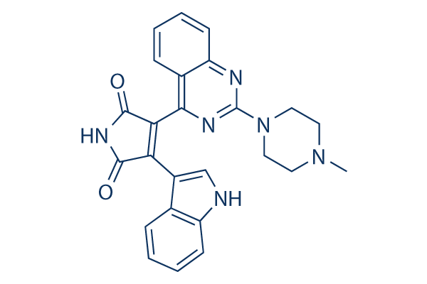
Chemische Struktur
Molekulargewicht: 438.48
Qualitätskontrolle
Zellkultur, Behandlung & Arbeitskonzentration
| Zelllinien | Assay-Typ | Konzentration | Inkubationszeit | Formulierung | Aktivitätsbeschreibung | PMID |
|---|---|---|---|---|---|---|
| T cell | Function Assay | 100 nM | 3 h | DMSO | inhibits rRNA synthesis | 25691158 |
| HUVECs | Function Assay | 500nM | 1 h | Reduces DTX-Triggered Endothelial Dysfunction | 25634538 | |
| A549 | Function Assay | 0.1 μM | 24 h | decreases the relative PKC-α level on cell membrane cotreated AS-IV | 25218161 | |
| A549 | Function Assay | 0.1 μM | 24 h | reduces the expression levels of MMP-2, MMP-9 and integrin β1 | 25218161 | |
| A549 | Growth Inhibition Assay | 0.1 μM | 24 h | enhances growth inhibition cotreated with AS-IV | 25218161 | |
| Mel202 | Growth Inhibition Assay | 0.5 μM | 3 h | DMSO | enhances IR-induced reduction in cell viability | 24595385 |
| 92.1 | Growth Inhibition Assay | 0.5 μM | 3 h | DMSO | enhances IR-induced reduction in cell viability | 24595385 |
| OCM3 | Growth Inhibition Assay | 0.5 μM | 3 h | DMSO | enhances IR-induced reduction in cell viability | 24595385 |
| Mel202 | Growth Inhibition Assay | 0.5 μM | 3 h | DMSO | increases IR-induced cell cycle arrest | 24595385 |
| 92.1 | Growth Inhibition Assay | 0.5 μM | 3 h | DMSO | increases IR-induced cell cycle arrest | 24595385 |
| OCM3 | Growth Inhibition Assay | 0.5 μM | 3 h | DMSO | increases IR-induced cell cycle arrest | 24595385 |
| Jeko-1 | Growth Inhibition Assay | 0-4 μM | DMSO | inhibits cell growth dose dependently | 24362935 | |
| Mino | Growth Inhibition Assay | 0-4 μM | DMSO | inhibits cell growth dose dependently | 24362935 | |
| Rec-1 | Growth Inhibition Assay | 0-4 μM | DMSO | inhibits cell growth dose dependently | 24362935 | |
| SP49 | Growth Inhibition Assay | 0-4 μM | DMSO | inhibits cell growth dose dependently | 24362935 | |
| Jeko-1 | Function Assay | 2.5 μM | 12 h | DMSO | downregulates NF-κB target genes | 24362935 |
| Mino | Function Assay | 2.5 μM | 12 h | DMSO | downregulates NF-κB target genes | 24362935 |
| Rec-1 | Function Assay | 2.5 μM | 12 h | DMSO | downregulates NF-κB target genes | 24362935 |
| SP49 | Function Assay | 2.5 μM | 12 h | DMSO | downregulates NF-κB target genes | 24362935 |
| CD3+ T | Function Assay | 0-500 nM | 1 h | inhibits NF-κB phosphorylation in a dose dependent manner | 23573283 | |
| Mel202 | Growth Inhibition Assay | 0-5 μM | 72 h | DMSO | inhibits cell growth dose dependently | 22653968 |
| Omm1.3 | Growth Inhibition Assay | 0-5 μM | 72 h | DMSO | inhibits cell growth dose dependently | 22653968 |
| 92.1 | Growth Inhibition Assay | 0-5 μM | 72 h | DMSO | inhibits cell growth dose dependently | 22653968 |
| Mel202 | Growth Inhibition Assay | 5 μM | 24 h | DMSO | induces G1 arrest | 22653968 |
| Omm1.3 | Growth Inhibition Assay | 5 μM | 24 h | DMSO | induces G1 arrest | 22653968 |
| 92.1 | Growth Inhibition Assay | 5 μM | 24 h | DMSO | induces G1 arrest | 22653968 |
| Mel202 | Apoptosis Assay | 5 μM | 72 h | DMSO | induces apoptosis slightly | 22653968 |
| Omm1.3 | Apoptosis Assay | 5 μM | 72 h | DMSO | induces apoptosis | 22653968 |
| 92.1 | Apoptosis Assay | 5 μM | 72 h | DMSO | induces apoptosis signifcantly | 22653968 |
| Mel202 | Function Assay | 5 μM | 24 h | inhibits expression and phosphorylation of PKC isoforms | 22653968 | |
| Omm1.3 | Function Assay | 5 μM | 24 h | inhibits expression and phosphorylation of PKC isoforms | 22653968 | |
| 92.1 | Function Assay | 5 μM | 24 h | inhibits expression and phosphorylation of PKC isoforms | 22653968 | |
| HBL1 | Growth Inhibition Assay | 0.16-20 μM | 5 d | IC50=0.5 μM | 21324920 | |
| TMD8 | Growth Inhibition Assay | 0.16-20 μM | 5 d | IC50=0.2 μM | 21324920 | |
| OCI-Ly10 | Growth Inhibition Assay | 0.16-20 μM | 5 d | IC50=1.3 μM | 21324920 | |
| U2932 | Growth Inhibition Assay | 0.16-20 μM | 5 d | IC50=10 μM | 21324920 | |
| OCI-Ly3 | Growth Inhibition Assay | 0.16-20 μM | 5 d | IC50>20 μM | 21324920 | |
| SuDHL2 | Growth Inhibition Assay | 0.16-20 μM | 5 d | IC50>20 μM | 21324920 | |
| SuDHL4 | Growth Inhibition Assay | 0.16-20 μM | 5 d | IC50>20 μM | 21324920 | |
| DB | Growth Inhibition Assay | 0.16-20 μM | 5 d | IC50>20 μM | 21324920 | |
| Jurkat IL-2 | Growth Inhibition Assay | IC50=6.71 ± 3.76 μM | 19940259 | |||
| PBMC IL-2 | Growth Inhibition Assay | IC50=4.84 ± 1.70 μM | 19940259 | |||
| Jurkat | Function assay | Inhibition of TCR/CD28-mediated human T cell activation in Jurkat cells expressing human IL2 promoter by luciferase reporter gene assay, IC50 = 0.054 μM. | 19827831 | |||
| Jurkat T | Function assay | 5 hrs | Inhibition of PKCtheta in human Jurkat T cells assessed as reduction in anti-CD3/CD28 antibody-induced T-cell activation by measuring decrease in IL-2 secretion after 5 hrs by luciferase reporter gene assay, IC50 = 0.081 μM. | 28131714 | ||
| B-cells | Function assay | Inhibition of PKCbeta in mouse B cells assessed as reduction in IgM-stimulated cell proliferation, IC50 = 0.234 μM. | 28131714 | |||
| bone marrow cells | Antiproliferative assay | 4 days | Antiproliferative activity against CBA mouse bone marrow cells assessed as inhibition of [3H]thymidine incorporation after 4 days, IC50 = 3.7 μM. | 19827831 | ||
| Klicken Sie hier, um weitere experimentelle Daten zu Zelllinien anzuzeigen | ||||||
Chemische Informationen, Lagerung & Stabilität
| Molekulargewicht | 438.48 | Formel | C25H22N6O2 |
Lagerung (Ab dem Eingangsdatum) | |
|---|---|---|---|---|---|
| CAS-Nr. | 425637-18-9 | SDF herunterladen | Lagerung von Stammlösungen |
|
|
| Synonyme | N/A | Smiles | CN1CCN(CC1)C2=NC3=CC=CC=C3C(=N2)C4=C(C(=O)NC4=O)C5=CNC6=CC=CC=C65 | ||
Löslichkeit
|
In vitro |
DMSO
: 87 mg/mL
(198.41 mM)
Ethanol : 40 mg/mL Water : Insoluble |
Molaritätsrechner
|
In vivo |
|||||
In-vivo-Formulierungsrechner (Klare Lösung)
Schritt 1: Geben Sie die untenstehenden Informationen ein (Empfohlen: Ein zusätzliches Tier zur Berücksichtigung von Verlusten während des Experiments)
Schritt 2: Geben Sie die In-vivo-Formulierung ein (Dies ist nur der Rechner, keine Formulierung. Bitte kontaktieren Sie uns zuerst, wenn es im Abschnitt "Löslichkeit" keine In-vivo-Formulierung gibt.)
Berechnungsergebnisse:
Arbeitskonzentration: mg/ml;
Methode zur Herstellung der DMSO-Stammlösung: mg Wirkstoff vorgelöst in μL DMSO ( Konzentration der Stammlösung mg/mL, Bitte kontaktieren Sie uns zuerst, wenn die Konzentration die DMSO-Löslichkeit der Wirkstoffcharge überschreitet. )
Methode zur Herstellung der In-vivo-Formulierung: Nehmen Sie μL DMSO Stammlösung, dann hinzufügenμL PEG300, mischen und klären, dann hinzufügenμL Tween 80, mischen und klären, dann hinzufügen μL ddH2O, mischen und klären.
Methode zur Herstellung der In-vivo-Formulierung: Nehmen Sie μL DMSO Stammlösung, dann hinzufügen μL Maisöl, mischen und klären.
Hinweis: 1. Bitte stellen Sie sicher, dass die Flüssigkeit klar ist, bevor Sie das nächste Lösungsmittel hinzufügen.
2. Achten Sie darauf, das/die Lösungsmittel der Reihe nach hinzuzufügen. Sie müssen sicherstellen, dass die bei der vorherigen Zugabe erhaltene Lösung eine klare Lösung ist, bevor Sie mit der Zugabe des nächsten Lösungsmittels fortfahren. Physikalische Methoden wie Vortex, Ultraschall oder ein heißes Wasserbad können zur Unterstützung des Lösens verwendet werden.
Wirkmechanismus
| Merkmale |
Unlike former PKC inhibitors, Sotrastaurin does not enhance apoptosis of murine T-cell blasts in a model of activation-induced cell death.
|
|---|---|
| Targets/IC50/Ki |
PKCθ
(Cell-free assay) 0.22 nM(Ki)
PKCβ1
(Cell-free assay) 0.64 nM(Ki)
PKCα
(Cell-free assay) 0.95 nM(Ki)
PKCη
(Cell-free assay) 1.8 nM(Ki)
PKCδ
(Cell-free assay) 2.1 nM(Ki)
PKCε
(Cell-free assay) 3.2 nM(Ki)
|
| In vitro |
Die Behandlung mit Sotrastaurin (AEB071) in Konzentrationen unter 10 μM hob in primären menschlichen und Maus-T-Zellen bei niedrigen nanomolaren Konzentrationen effektiv Marker der frühen T-Zell-Aktivierung – wie Interleukin-2-Sekretion und CD25-Expression – auf. Bei 200 nM hemmt es die CD3/CD28-Antikörper- und Alloantigen-induzierte T-Zell-Proliferation ohne unspezifische antiproliferative Effekte. Darüber hinaus beeinträchtigt diese Verbindung (<3 μM) die Lymphozytenfunktion-assoziierte Antigen-1-vermittelte T-Zell-Adhäsion erheblich. In Konzentrationen unter 20 μM beeinträchtigt es selektiv die Proliferation von CD79-mutierten ABC DLBCL-Zelllinien, korrelierend mit einer reduzierten NF-κB-Signalaktivität. Eine Konzentration von 5 μM induziert G1-Arrest und/oder Zelltod in CD79-mutierten Zellen. |
| Kinase-Assay |
Proteinkinase-Assays
|
|
Sotrastaurin (AEB071) wurde auf klassische und neue PKC-Isotypen unter Verwendung der Szintillations-Proximity-Assay-Technologie getestet. Kurz gesagt, der Assay wird in 20 mM Tris-HCl-Puffer, pH 7,4, und 0,1 % Rinderserumalbumin durchgeführt, indem 1,5 μM des Peptidsubstrats mit 10 μM [33P]ATP, 10 mM Mg (NO3)2, 0,2 mM CaCl2 und PKC in einer Proteinkonzentration von 25 bis 400 ng/ml inkubiert wird, sowie Lipidvesikel, die 30 Mol-% Phosphatidylserin, 5 Mol-% Diacylglycerol (DAG) und 65 Mol-% Phosphatidylcholin bei einer finalen Lipidkonzentration von 0,5 μM enthalten. Die Inkubation erfolgt 60 min bei Raumtemperatur. Die Reaktion wird durch Zugabe von 50 μl einer Mischung gestoppt, die 100 mM EDTA, 200 μM ATP, 0,1 % Triton X-100 und 0,375 μg/Well Streptavidin-beschichtete Szintillations-Proximity-Assay-Beads in PBS ohne Ca2+ und Mg2+ enthält. Die inkorporierte Radioaktivität wird in einem MicroBetaTrilux-Zähler für 1 min gemessen.
|
|
| In vivo |
In einem subkutanen TMD8-Xenograftmodell an SCID-Mäusen führt Sotrastaurin (AEB071) (80 mg/kg) zu einer signifikanten Hemmung des in-vivo-Tumorwachstums. Bei oraler Verabreichung von 10 mg/kg und 30 mg/kg zweimal täglich zeigt diese Verbindung eine dosisabhängige immunsuppressive Wirkung, die zu einer ausgeprägten Verlängerung des Überlebens von Herzallotransplantaten bei Ratten führt. |
Literatur |
|
Anwendungen
| Methoden | Biomarker | Bilder | PMID |
|---|---|---|---|
| Western blot | p-Marcks / p-ERK / p-AKT / p-S6 / Marcks / ERK / AKT / S6 PKCα / PKCδ / PKCβ / PKCε / PKCθ Bcl-xl / XIAP / Survivin Cyclin D1 / p27(Kip1) pPKCδ/θ / phosphorylated MARCKS / p53 / MDM2 / PUMA / p21 |
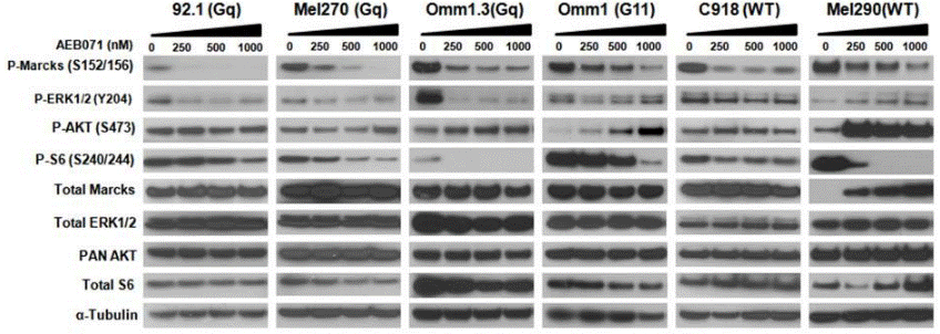
|
22653968 |
| Growth inhibition assay | Cell viability |
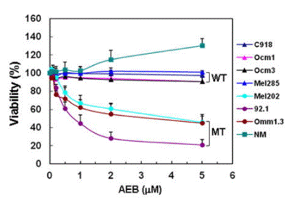
|
22653968 |
Klinische Studieninformationen
(Daten von https://clinicaltrials.gov, aktualisiert am 2024-05-22)
| NCT-Nummer | Rekrutierung | Erkrankungen | Sponsor/Kooperationspartner | Startdatum | Phasen |
|---|---|---|---|---|---|
| NCT02273219 | Completed | Uveal Melanoma |
Columbia University |
November 19 2014 | Phase 1 |
| NCT01801358 | Terminated | Uveal Melanoma |
Array Biopharma now a wholly owned subsidiary of Pfizer|Array BioPharma |
August 2013 | Phase 1|Phase 2 |
| NCT01430416 | Completed | Uveal Melanoma |
Novartis Pharmaceuticals|Novartis |
December 20 2011 | Phase 1 |
| NCT01402440 | Terminated | Diffuse Large B-Cell Lymphoma |
Novartis Pharmaceuticals|Novartis |
November 2011 | Phase 1 |
Technischer Support
Tel: +1-832-582-8158 Ext:3
Wenn Sie weitere Fragen haben, hinterlassen Sie bitte eine Nachricht.
Häufig gestellte Fragen
Frage 1:
Could you give me the information about how to prepare it for oral administration in mice?
Antwort:
It can be dissolved in 2% DMSO/30% PEG 300/ddH2O at 10 mg/ml as a clear solution which can be used for injection, and in 2% DMSO/corn oil at 10 mg/ml as a suspension for oral administration.
Produkte sind nur für Forschungszwecke bestimmt. Nicht für den menschlichen Gebrauch. Wir verkaufen nicht an Patienten.
©Copyright 2013 Selleck Chemicals. Alle Rechte vorbehalten.