nur für Forschungszwecke
Stattic STAT3-Inhibitor
Kat.-Nr.S7024
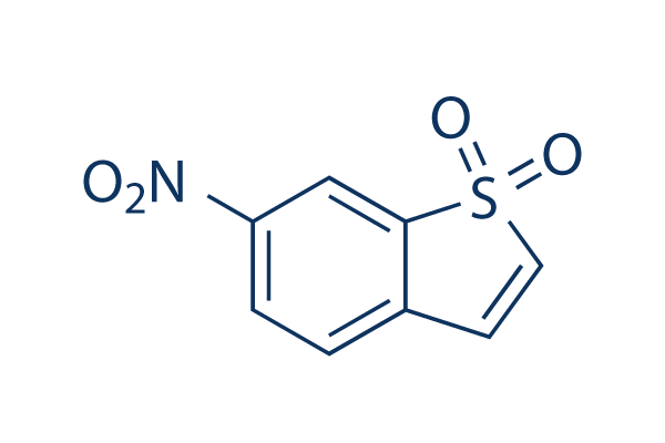
Chemische Struktur
Molekulargewicht: 211.19
Qualitätskontrolle
| Verwandte Ziele | EGFR JAK Pim |
|---|---|
| Weitere STAT Inhibitoren | Napabucasin (BBI608) NSC 74859 (S3I-201) Cryptotanshinone (Tanshinone C) C188-9 (TTI-101) SH-4-54 BP-1-102 AS1517499 Nifuroxazide HO-3867 Homoharringtonine (HHT) |
Zellkultur, Behandlung & Arbeitskonzentration
| Zelllinien | Assay-Typ | Konzentration | Inkubationszeit | Formulierung | Aktivitätsbeschreibung | PMID |
|---|---|---|---|---|---|---|
| H9c2 | Function Assay | 10 μM | 4 h | reverses the effects of IL-27 | 26339633 | |
| NPC | Function Assay | 0-7.5 µM | abolishes EMT-like molecular alterations, and cell migration and invasion induced by RKIP knockdown | 25915430 | ||
| HASMC | Function Assay | 1.25-5 μM | 20 min | DMSO | inhibits p-(Y)-STAT-1,3,5 signals | 25849622 |
| H9c2 | Function Assay | 2/10 μM | 2 h | DMSO | abrogates the cytoprotective effects of IL-27 against SH | 25820907 |
| A431 | Growth Inhibition Assay | 2 μM | 2 h | blocks EGF-reversed decreases in cell viability | 25720435 | |
| A431 | Growth Inhibition Assay | 2 μM | 2 h | increases in apoptosis induced by shikonin | 25720435 | |
| SiHa | Cell Viability Assay | 5-75 nM | 24 h | shows morphology of a typical apoptotic cell and dose-dependent loss of cell viability | 25539644 | |
| SiHa | Function Assay | 5-75 nM | 24 h | reduces the phosphorylation at the tyrosine residue 705 | 25539644 | |
| ECA109 | Growth Inhibition Assay | 0-20 μM | 24 h | IC50=5.50 μM | 25492480 | |
| TE13 | Growth Inhibition Assay | 0-20 μM | 24 h | IC50=6.15 μM | 25492480 | |
| KYSE150 | Growth Inhibition Assay | 0-20 μM | 24 h | IC50=12.64 μM | 25492480 | |
| ECA109 | Clonogenic Survival Assay | 0.5 μM | 24 h | suppresses the clonogenic formation | 25492480 | |
| TE13 | Clonogenic Survival Assay | 0.5 μM | 24 h | suppresses the clonogenic formation | 25492480 | |
| KYSE150 | Clonogenic Survival Assay | 0.5 μM | 24 h | suppresses the clonogenic formation | 25492480 | |
| ECA109 | Function Assay | 0.5 μM | 24 h | enhances IR-induced generation of DSBs | 25492480 | |
| PC3M-1E8 | Function Assay | 2.5/5/10 μM | 0-4 h | inhibits the STAT3 activation in a dose- and time-dependent manner | 25261365 | |
| PC3M-1E8 | Function Assay | 10 μM | 24 h | downregulates Bcl-xL, survivin and c-Myc | 25261365 | |
| PC3M-1E8 | Function Assay | 10 μM | 24 h | inhibits IL-6 induced STAT3 activation and the IL-6-induced STAT3 activation | 25261365 | |
| PC3M-1E8 | Clonogenic Survival Assay | 2.5/5/10 μM | inhibits the colony formation significantly | 25261365 | ||
| MDA-MB-231 | Function Assay | 20 μM | 2 h | exhibits Snail and E-cadherin expression | 25153349 | |
| H9c2 | Function Assay | 20 µM | 30 min | DMSO | abolishes propofol-induced AKT phosphorylation at both ser473 and thr308 | 25105067 |
| HaCaT | Growth Inhibition Assay | 10 µM | 20 min | DMSO | enhances sorafenib- and sunitinib-induced growth inhibition | 25013907 |
| Caki-1 | Growth Inhibition Assay | 10 µM | 20 min | DMSO | enhances sorafenib- and sunitinib-induced growth inhibition | 25013907 |
| HaCaT | Apoptosis Assay | 10 µM | 20 min | DMSO | increases proportions of apoptotic cells due to treatment with sorafenib or sunitinib | 25013907 |
| FHL-primed hNSCs | Cell Viability Assay | 0.02-5 μM | 72 h | leads to the loss of cell viability at high concentration | 24945434 | |
| ELL-primed hNSCs | Cell Viability Assay | 0.02-5 μM | 72 h | leads to the loss of cell viability at high concentration | 24945434 | |
| SS | Cell Viability Assay | 1-10 μM | 72 h | DMSO | causes a dose-dependent inhibition of the viability | 24756111 |
| SeAx | Cell Viability Assay | 1-10 μM | 72 h | DMSO | causes a dose-dependent inhibition of the viability | 24756111 |
| HuT-78 | Cell Viability Assay | 1-10 μM | 72 h | DMSO | causes a dose-dependent inhibition of the viability | 24756111 |
| CD4+ | Apoptosis Assay | 10 μm | 24 h | DMSO | induces apoptosis strongly | 24756111 |
| MCF-7 | Growth Inhibition Assay | 0.469-3.75 μM | 5 d | reduces cell number significantly | 24728078 | |
| MCF-7/LCC1 | Growth Inhibition Assay | 0.469-3.75 μM | 5 d | reduces cell number significantly | 24728078 | |
| MCF-7/LCC9 | Growth Inhibition Assay | 0.469-3.75 μM | 5 d | reduces cell number significantly | 24728078 | |
| HaCaT | Growth Inhibition Assay | 10 µM | 20 min | DMSO | enhances everolimus-induced cell growth inhibition | 24423131 |
| HaCaT | Apoptosis Assay | 10 µM | 20 min | DMSO | enhances the apoptotic effects of everolimus | 24423131 |
| MDA-MB-231 | Function Assay | 10 µM | 24 h | DMSO | reduces P-STAT3 expression | 24376586 |
| SUM-159 | Function Assay | 10 µM | 24 h | DMSO | reduces P-STAT3 expression | 24376586 |
| SK-BR-3 | Function Assay | 10 µM | 24 h | DMSO | reduces P-STAT3 expression | 24376586 |
| MCF7-HER2 | Growth Inhibition Assay | 0-10 μM | 48 h | DMSO | induces cell death dose dependently | 24297508 |
| MCF7-HER2 | Function Assay | 5 μM | 24 h | DMSO | diminishes Sox-2, Oct-4, and slug expression | 24297508 |
| MCF7-HER2 | Function Assay | 5 μM | 24 h | DMSO | decreases the expression levels of EMT markers, vimentin and slug | 24297508 |
| MCF7-HER2 | Growth Inhibition Assay | 5 μM | 24 h | DMSO | enhances cell growth inhibition combined with Herceptin | 24297508 |
| HMECs | Function Assay | 10 μM | 2 h | inhibits IFNα mediated phosphorylation of STAT1, STAT2 and STAT3 | 24211327 | |
| HTR8/SVneo | Function Assay | 1 μM | 1 h | suppressed OSM-induced STAT3 phosphorylation | 24060241 | |
| HTR8/SVneo | Function Assay | 0.5/1 μM | 48 h | restores the expression of E-cadherin suppressed by OSM | 24060241 | |
| HTR8/SVneo | Function Assay | 1 μM | 48 h | significantly increases migration by OSM | 24060241 | |
| C13* | Apoptosis Assay | 0-10 μM | 24/48 h | induces apoptosis in a dose and time dependent manner | 23962558 | |
| OV2008 | Apoptosis Assay | 0-10 μM | 24/48 h | induces apoptosis in a dose and time dependent manner | 23962558 | |
| C13* | Apoptosis Assay | 24/48 h | enhances cisplatin-induced apoptosis | 23962558 | ||
| OV2008 | Apoptosis Assay | 24/48 h | enhances cisplatin-induced apoptosis | 23962558 | ||
| W480 | Function Assay | 2.5/10 μM | 30 min | DMSO | sensitizes cells to chemoradiotherapy in a dose-dependent manner | 23934972 |
| SW837 | Function Assay | 2.5/10 μM | 30 min | DMSO | sensitizes cells to chemoradiotherapy in a dose-dependent manner | 23934972 |
| T24 | Function Assay | 2/10/20 μM | 24 h | causes dose-dependent inhibition of the CXCL12-induced increase of invading cells | 23526079 | |
| CNE1 | Function Assay | 20 µM | 48 h | blocks the IL-6 increased phosphorylation of Stat3 | 23382914 | |
| CNE2 | Function Assay | 20 µM | 48 h | blocks the IL-6 increased phosphorylation of Stat3 | 23382914 | |
| HONE1 | Function Assay | 20 µM | 48 h | blocks the IL-6 increased phosphorylation of Stat3 | 23382914 | |
| CNE1 | Growth Inhibition Assay | 4 μM | significantly reduces cell viability | 23382914 | ||
| CNE1 | Function Assay | 0-20 μM | 0-4 h | inhibits Stat3 activation in a dose- and time-dependent manner | 23382914 | |
| CNE2 | Function Assay | 0-20 μM | 0-4 h | inhibits Stat3 activation in a dose- and time-dependent manner | 23382914 | |
| HONE1 | Function Assay | 0-20 μM | 0-4 h | inhibits Stat3 activation in a dose- and time-dependent manner | 23382914 | |
| CNE1 | Cell Viability Assay | 0.5-64 μM | 48 h | suppresses cell viability in a dose- and time-dependent manner | 23382914 | |
| CNE2 | Cell Viability Assay | 0.5-64 μM | 48 h | suppresses cell viability in a dose- and time-dependent manner | 23382914 | |
| HONE1 | Cell Viability Assay | 0.5-64 μM | 48 h | suppresses cell viability in a dose- and time-dependent manner | 23382914 | |
| C666-1 | Cell Viability Assay | 0.5-64 μM | 48 h | suppresses cell viability in a dose- and time-dependent manner | 23382914 | |
| CNE1 | Apoptosis Assay | 10 µM | 48 h | induces apoptosis | 23382914 | |
| CNE2 | Apoptosis Assay | 10 µM | 48 h | induces apoptosis | 23382914 | |
| HONE1 | Apoptosis Assay | 10 µM | 48 h | induces apoptosis | 23382914 | |
| CNE2 | Cell Viability Assay | 1/2 μM | 48 h | sensitize cells to radiotherapy | 23382914 | |
| HONE1 | Cell Viability Assay | 1/2 μM | 48 h | sensitize cells to radiotherapy | 23382914 | |
| C666-1 | Cell Viability Assay | 1/2 μM | 48 h | sensitize cells to radiotherapy | 23382914 | |
| HEC-1A | Function Assay | 1 μM | 24 h | DMSO | blocks the MUC20-enhanced invasion triggered by 10% FBS | 23262208 |
| RL95-2 | Function Assay | 1 μM | 24 h | DMSO | blocks the MUC20-enhanced invasion triggered by 10% FBS | 23262208 |
| HEC-1A | Function Assay | 1 μM | 24 h | DMSO | blocks the MUC20-enhanced invasion triggered by EGF | 23262208 |
| RL95-2 | Function Assay | 1 μM | 24 h | DMSO | blocks the MUC20-enhanced invasion triggered by EGF | 23262208 |
| CT26 | Function Assay | 20 mM | 1 h | suppresses HGF-induced VEGF expression | 23233163 | |
| UM-SCC-17B | Growth Inhibition Assay | IC50=2.562 ± 0.409 μM, GI50=1.279 ± 0.194 μM | 22770899 | |||
| OSC-19 | Growth Inhibition Assay | IC50=3.481 ± 0.953 μM, GI50=1.366 ± 0.770 μM | 22770899 | |||
| Cal33 | Growth Inhibition Assay | IC50=2.282 ± 0.423 μM, GI50=1.349 ± 0.363 μM | 22770899 | |||
| UM-SCC-22B | Growth Inhibition Assay | IC50=2.648 ± 0.542 μM, GI50=1.320 ± 0.204 μM | 22770899 | |||
| UM-SCC-17B | Function Assay | 0-30 μM | 0-24 h | inhibits STAT3 activation dose and time dependently | 22770899 | |
| OSC-19 | Function Assay | 0-30 μM | 0-24 h | inhibits STAT3 activation dose and time dependently | 22770899 | |
| Cal33 | Function Assay | 0-30 μM | 0-24 h | inhibits STAT3 activation dose and time dependently | 22770899 | |
| UM-SCC-22B | Function Assay | 0-30 μM | 0-24 h | inhibits STAT3 activation dose and time dependently | 22770899 | |
| U-87MG | Cell Viability Assay | 0-10 μM | 72 h | DMSO | inhibits cell viability dose dependently | 25436682 |
| U-373MG | Cell Viability Assay | 0-10 μM | 72 h | DMSO | inhibits cell viability dose dependently | 25436682 |
| SH-SY5Y | Cell Viability Assay | 0-10 μM | 72 h | DMSO | inhibits cell viability dose dependently | 25436682 |
| Tu-9648 | Cell Viability Assay | 0-10 μM | 72 h | DMSO | inhibits cell viability dose dependently | 25436682 |
| Neuro-2a | Cell Viability Assay | 0-10 μM | 72 h | DMSO | inhibits cell viability dose dependently | 25436682 |
| PCNs | Cell Viability Assay | 0-10 μM | 72 h | DMSO | inhibits cell viability dose dependently | 25436682 |
| PGCs | Cell Viability Assay | 0-10 μM | 72 h | DMSO | inhibits cell viability dose dependently | 25436682 |
| RAW264.7 | Function Assay | 10 μM | 12 h | abrogates the mRNA expressions of JAK2, STAT1, STAT2, and STAT3 induced by DON and T-2 toxin | 22454431 | |
| RAW264.7 | Apoptosis Assay | 5/10 μM | 45 min | enhances toxins induced apoptosis and MMP loss | 22454431 | |
| SW480 | Cell Viability Assay | 5/10/20 μM | 72 h | inhibits cell viability of the ALDH+/CD133+ cells | 21900397 | |
| HCT116 | Cell Viability Assay | 5/10/20 μM | 72 h | inhibits cell viability of the ALDH+/CD133+ cells | 21900397 | |
| DLD-1 | Cell Viability Assay | 5/10/20 μM | 72 h | inhibits cell viability of the ALDH+/CD133+ cells | 21900397 | |
| SNU387 | Cell Viability Assay | 20 μM | 24 h | reduces cell viability | 21311975 | |
| SNU398 | Cell Viability Assay | 20 μM | 24 h | reduces cell viability | 21311975 | |
| HepG2 | Cell Viability Assay | 20 μM | 24 h | reduces cell viability | 21311975 | |
| Huh-7 | Cell Viability Assay | 20 μM | 24 h | reduces cell viability | 21311975 | |
| VSMC | Growth Inhibition Assay | 3/5/10 μM | 30 min | DMSO | prevents PDGF- and thrombin-mediated VSMC proliferation in a dose-dependent manner | 20847306 |
| MDA-MB-231 | Apoptosis Assay | 10 μM | 24 h | DMSO | induces apoptosis | 17114005 |
| MDA-MB-435S | Apoptosis Assay | 10 μM | 24 h | DMSO | induces apoptosis | 17114005 |
| AsPC1 | Antiproliferative assay | 72 hrs | Antiproliferative activity against human AsPC1 cells assessed as inhibition of cell proliferation after 72 hrs by MTS assay, IC50 = 1.32 μM. | 24904966 | ||
| MDA-MB-231 | Antiproliferative assay | 72 hrs | Antiproliferative activity against ER-negative and triple-negative human MDA-MB-231 cells assessed as inhibition of cell proliferation after 72 hrs by MTS assay, IC50 = 2.89 μM. | 24904966 | ||
| MCF7 | Antiproliferative assay | 72 hrs | Antiproliferative activity against ER-positive human MCF7 cells assessed as inhibition of cell proliferation after 72 hrs by MTS assay, IC50 = 3.6 μM. | 24904966 | ||
| PANC1 | Antiproliferative assay | 72 hrs | Antiproliferative activity against human PANC1 cells assessed as inhibition of cell proliferation after 72 hrs by MTS assay, IC50 = 3.77 μM. | 24904966 | ||
| MDA-MB-231 | Cytotoxicity assay | 48 hrs | Cytotoxicity against human MDA-MB-231 cells assessed as growth inhibition after 48 hrs by MTT assay, IC50 = 1.56 μM. | 26396689 | ||
| MDA-MB-435S | Cytotoxicity assay | 48 hrs | Cytotoxicity against human MDA-MB-435S cells assessed as growth inhibition after 48 hrs by MTT assay, IC50 = 1.87 μM. | 26396689 | ||
| MCF7 | Cytotoxicity assay | 48 hrs | Cytotoxicity against human MCF7 cells assessed as growth inhibition after 48 hrs by MTT assay, IC50 = 2.16 μM. | 26396689 | ||
| A549 | Cytotoxicity assay | 48 hrs | Cytotoxicity against human A549 cells assessed as growth inhibition after 48 hrs by MTT assay, IC50 = 2.5 μM. | 26396689 | ||
| DU145 | Cytotoxicity assay | 48 hrs | Cytotoxicity against human DU145 cells assessed as growth inhibition after 48 hrs by MTT assay, IC50 = 2.5 μM. | 26396689 | ||
| PANC1 | Cytotoxicity assay | 48 hrs | Cytotoxicity against human PANC1 cells assessed as growth inhibition after 48 hrs by MTT assay, IC50 = 2.9 μM. | 26396689 | ||
| HCT116 | Antiproliferative assay | 72 hrs | Antiproliferative activity against human HCT116 cells after 72 hrs by MTT assay, IC50 = 1.08 μM. | 27718470 | ||
| MDA-MB-231 | Antiproliferative assay | 72 hrs | Antiproliferative activity against human MDA-MB-231 cells after 72 hrs by MTT assay, IC50 = 1.68 μM. | 27718470 | ||
| MCF7 | Antiproliferative assay | 72 hrs | Antiproliferative activity against human MCF7 cells after 72 hrs by MTT assay, IC50 = 2.36 μM. | 27718470 | ||
| A549 | Antiproliferative assay | 72 hrs | Antiproliferative activity against human A549 cells after 72 hrs by MTT assay, IC50 = 4.4 μM. | 27718470 | ||
| AD293 | Function assay | 6 hrs | Inhibition of IFNgamma-stimulated GFP/FLAG-tagged STAT3 dimerization in human AD293 cells incubated for 6 hrs by Western blot analysis, IC50 = 5.1 μM. | 30228000 | ||
| MDA-MB-231 | Function assay | 1 to 10 uM | 12 hrs | Inhibition of STAT3 phosphorylation at Tyr705 in human MDA-MB-231 cells at 1 to 10 uM after 12 hrs by western blot analysis | 24904966 | |
| MDA-MB-231 | Anticancer assay | 1 to 10 uM | 48 hrs | Anticancer activity against human MDA-MB-231 cells assessed as cell growth inhibition, apoptosis and cellular morphological changes at 1 to 10 uM after 48 hrs by light microscopy | 24904966 | |
| MDA-MB-231 | Function assay | 1 to 10 uM | 12 hrs | Decrease in STAT3 protein expression in human MDA-MB-231 cells at 1 to 10 uM after 12 hrs by western blot analysis | 24904966 | |
| MCF7 | Function assay | 12 hrs | Inhibition of STAT3 phosphorylation at Y705 in human MCF7 cells after 12 hrs by Western blot analysis | 26396689 | ||
| MDA-MB-435S | Function assay | 12 hrs | Inhibition of STAT3 phosphorylation at Y705 in human MDA-MB-435S cells after 12 hrs by Western blot analysis | 26396689 | ||
| MDA-MB-231 | Function assay | 12 hrs | Inhibition of STAT3 phosphorylation at Y705 in human MDA-MB-231 cells after 12 hrs by Western blot analysis | 26396689 | ||
| Klicken Sie hier, um weitere experimentelle Daten zu Zelllinien anzuzeigen | ||||||
Chemische Informationen, Lagerung & Stabilität
| Molekulargewicht | 211.19 | Formel | C8H5NO4S |
Lagerung (Ab dem Eingangsdatum) | |
|---|---|---|---|---|---|
| CAS-Nr. | 19983-44-9 | SDF herunterladen | Lagerung von Stammlösungen |
|
|
| Synonyme | N/A | Smiles | C1=CC(=CC2=C1C=CS2(=O)=O)[N+](=O)[O-] | ||
Löslichkeit
|
In vitro |
DMSO
: 42 mg/mL
(198.87 mM)
Water : Insoluble Ethanol : Insoluble |
Molaritätsrechner
|
In vivo |
|||||
In-vivo-Formulierungsrechner (Klare Lösung)
Schritt 1: Geben Sie die untenstehenden Informationen ein (Empfohlen: Ein zusätzliches Tier zur Berücksichtigung von Verlusten während des Experiments)
Schritt 2: Geben Sie die In-vivo-Formulierung ein (Dies ist nur der Rechner, keine Formulierung. Bitte kontaktieren Sie uns zuerst, wenn es im Abschnitt "Löslichkeit" keine In-vivo-Formulierung gibt.)
Berechnungsergebnisse:
Arbeitskonzentration: mg/ml;
Methode zur Herstellung der DMSO-Stammlösung: mg Wirkstoff vorgelöst in μL DMSO ( Konzentration der Stammlösung mg/mL, Bitte kontaktieren Sie uns zuerst, wenn die Konzentration die DMSO-Löslichkeit der Wirkstoffcharge überschreitet. )
Methode zur Herstellung der In-vivo-Formulierung: Nehmen Sie μL DMSO Stammlösung, dann hinzufügenμL PEG300, mischen und klären, dann hinzufügenμL Tween 80, mischen und klären, dann hinzufügen μL ddH2O, mischen und klären.
Methode zur Herstellung der In-vivo-Formulierung: Nehmen Sie μL DMSO Stammlösung, dann hinzufügen μL Maisöl, mischen und klären.
Hinweis: 1. Bitte stellen Sie sicher, dass die Flüssigkeit klar ist, bevor Sie das nächste Lösungsmittel hinzufügen.
2. Achten Sie darauf, das/die Lösungsmittel der Reihe nach hinzuzufügen. Sie müssen sicherstellen, dass die bei der vorherigen Zugabe erhaltene Lösung eine klare Lösung ist, bevor Sie mit der Zugabe des nächsten Lösungsmittels fortfahren. Physikalische Methoden wie Vortex, Ultraschall oder ein heißes Wasserbad können zur Unterstützung des Lösens verwendet werden.
Wirkmechanismus
| Merkmale |
Stattic is the first non-peptide small molecule with inhibitory activity against STAT3 SH2 domain regardless of the STAT3 phosphorylation state in vitro.
|
|---|---|
| Targets/IC50/Ki |
STAT3
(Cell-free assay) 5.1 μM
|
| In vitro |
Stattic hemmt die Bindung eines Phosphotyrosin-haltigen Peptids, das vom gp130-Rezeptor stammt, an die STAT3 SH2-Domäne auf eine stark temperaturabhängige Weise. Diese Verbindung hat nur eine sehr schwache Wirkung auf die Bindung eines Tyrosin-phosphorylierten Peptids an die SH2-Domäne der Tyrosinkinase Lck. Und sie hemmt nicht die Dimerisierung von zwei anderen dimeren Transkriptionsfaktoren (c-Myc/Max und Jun/Jun). Sie hemmt auch fluoresceinmarkierte Phosphopeptide an die SH2-Domänen von STAT1 und STAT5b. Diese Chemikalie hemmt selektiv die DNA-Bindung von STAT3-Homodimeren bei einer Konzentration von 10 μM. Es wurde gezeigt, dass sie die zelluläre Phosphorylierung von STAT3 an Tyr705 hemmt, mit geringem Einfluss auf die STAT1-Phosphorylierung an Tyr701 (in HepG2-Zellen) oder die Phosphorylierung von JAK1, JAK2 und c-Src (in MDA-MB-231- und MDA-MB-235S-Zellen). Diese Verbindung erhöht die Apoptose-Rate von STAT3-abhängigen Brustkrebszelllinien. |
| Kinase-Assay |
Hochdurchsatz-Screening und Fluoreszenzpolarisationstests
|
|
Das Screening wird bei etwa 30 °C durchgeführt. Die Spezifität der Screening-Treffer wird in analogen Assays zur Bindung der Testverbindungen an die SH2-Domänen von STAT1, STAT5 und Lck validiert. Die Endkonzentration der Pufferkomponenten, die für alle FP-Assays verwendet werden, beträgt 10 mM HEPES (pH 7,5), 1 mM EDTA, 0,1 % Nonidet P-40, 50 mM NaCl und 10 % DMSO. Die Abwesenheit von Dithiothreitol ist für die inhibitorische Aktivität essentiell. Die Sequenzen der Peptide sind: STAT3, 5-Carboxyfluorescein-GY(PO3H2)LPQTV-NH2; STAT1, 5-Carboxyfluorescein-GY(PO3H2)DKPHVL; STAT5, 5-Carboxyfluorescein-GY(PO3H2)LVLDKW; und Lck, 5-Carboxyfluorescein-GY(PO3H2)EEIP. Für die Spezifitätsanalyse bei 30 °C werden Proteine in einer Konzentration von 150 nM (STAT1, STAT3 und STAT5) verwendet. Für die Spezifitätsanalyse bei 37 °C werden Proteine in einer Konzentration von 370 nM (STAT3) oder 100 nM (Lck) verwendet. Proteine werden mit Testverbindungen in Eppendorf-Röhrchen bei den angegebenen Temperaturen für 60 Minuten inkubiert, bevor die entsprechenden 5-Carboxyfluorescein-markierten Peptide (Endkonzentration: 10 nM) hinzugefügt werden. Vor der Messung bei Raumtemperatur lässt man die Mischungen mindestens 30 Minuten lang äquilibrieren. Testverbindungen werden in den angegebenen Konzentrationen verwendet, verdünnt aus einem 20-fachen Stammlösung in DMSO. Bindungskurven und Hemmkurven werden mit SigmaPlot angepasst. Alle Kompetitionskurven werden dreimal in unabhängigen Experimenten wiederholt.
|
Literatur |
|
Anwendungen
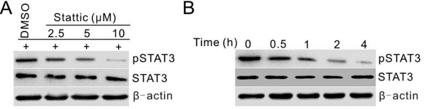
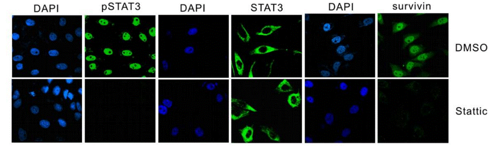
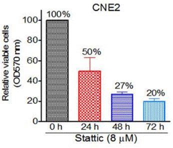
| Methoden | Biomarker | Bilder | PMID |
|---|---|---|---|
| Western blot | p-STAT3 / STAT3 Survivin / c-Myc / Bcl-xl PARP / C-PARP / Caspase-3 / C-Caspse-3 |
|
25261365 |
| Immunofluorescence | p-STAT3 / STAT3 / Survivin |
|
25261365 |
| Growth inhibition assay | Cell viability |
|
23382914 |
| ELISA | BDNF |
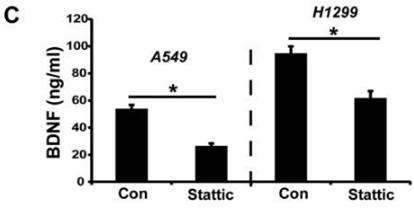
|
27456333 |
Technischer Support
Tel: +1-832-582-8158 Ext:3
Wenn Sie weitere Fragen haben, hinterlassen Sie bitte eine Nachricht.
Produkte sind nur für Forschungszwecke bestimmt. Nicht für den menschlichen Gebrauch. Wir verkaufen nicht an Patienten.
©Copyright 2013 Selleck Chemicals. Alle Rechte vorbehalten.