
nur für Forschungszwecke
Latrepirdine 2HCl GluR Antagonist
Kat.-Nr.S1245
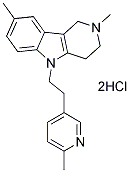
Chemische Struktur
Molekulargewicht: 392.37
Qualitätskontrolle
Chemische Informationen, Lagerung & Stabilität
| Molekulargewicht | 392.37 | Formel | C21H25N3.2HCl |
Lagerung (Ab dem Eingangsdatum) | |
|---|---|---|---|---|---|
| CAS-Nr. | 97657-92-6 | -- | Lagerung von Stammlösungen |
|
|
| Synonyme | N/A | Smiles | CC1=CC2=C(C=C1)N(C3=C2CN(CC3)C)CCC4=CN=C(C=C4)C.Cl.Cl | ||
Löslichkeit
|
In vitro |
DMSO
: 78 mg/mL
(198.79 mM)
Ethanol : 26 mg/mL Water : Insoluble |
Molaritätsrechner
|
In vivo |
|||||
In-vivo-Formulierungsrechner (Klare Lösung)
Schritt 1: Geben Sie die untenstehenden Informationen ein (Empfohlen: Ein zusätzliches Tier zur Berücksichtigung von Verlusten während des Experiments)
Schritt 2: Geben Sie die In-vivo-Formulierung ein (Dies ist nur der Rechner, keine Formulierung. Bitte kontaktieren Sie uns zuerst, wenn es im Abschnitt "Löslichkeit" keine In-vivo-Formulierung gibt.)
Berechnungsergebnisse:
Arbeitskonzentration: mg/ml;
Methode zur Herstellung der DMSO-Stammlösung: mg Wirkstoff vorgelöst in μL DMSO ( Konzentration der Stammlösung mg/mL, Bitte kontaktieren Sie uns zuerst, wenn die Konzentration die DMSO-Löslichkeit der Wirkstoffcharge überschreitet. )
Methode zur Herstellung der In-vivo-Formulierung: Nehmen Sie μL DMSO Stammlösung, dann hinzufügenμL PEG300, mischen und klären, dann hinzufügenμL Tween 80, mischen und klären, dann hinzufügen μL ddH2O, mischen und klären.
Methode zur Herstellung der In-vivo-Formulierung: Nehmen Sie μL DMSO Stammlösung, dann hinzufügen μL Maisöl, mischen und klären.
Hinweis: 1. Bitte stellen Sie sicher, dass die Flüssigkeit klar ist, bevor Sie das nächste Lösungsmittel hinzufügen.
2. Achten Sie darauf, das/die Lösungsmittel der Reihe nach hinzuzufügen. Sie müssen sicherstellen, dass die bei der vorherigen Zugabe erhaltene Lösung eine klare Lösung ist, bevor Sie mit der Zugabe des nächsten Lösungsmittels fortfahren. Physikalische Methoden wie Vortex, Ultraschall oder ein heißes Wasserbad können zur Unterstützung des Lösens verwendet werden.
Wirkmechanismus
| Targets/IC50/Ki |
GluR
Histamine receptor
5-HT
|
|---|---|
| In vitro |
Latrepirdine erhöht die Succinatdehydrogenase-Aktivität (MTT-Assay), das mitochondriale Membranpotenzial (DeltaPsim) und die zellulären ATP-Spiegel in primären Maus-Kortikalneuronen und menschlichen Neuroblastomzellen (SH-SY5Y). Latrepirdine verbessert die mitochondriale Funktion sowohl in Abwesenheit als auch in Anwesenheit von Stress, und mit Dimebon behandelte Zellen sind teilweise geschützt, um die Zellviabilität aufrechtzuerhalten. Latrepirdine führt zu einer verstärkten mTOR- und Atg5-abhängigen Autophagie in kultivierten Säugetierzellen. Latrepirdine stimuliert die MTOR- und ATG5-abhängige Autophagie, was zu einer Reduzierung der intrazellulären Spiegel von APP-Metaboliten, einschließlich Aβ, in kultivierten Zellen führt. Latrepirdine stimuliert den Abbau von α-syn in differenzierten SH-SY5Y-Neuronen und im Gehirn von Mäusen nach chronischer Verabreichung, parallel zur Erhöhung der Spiegel von Markern der autophagischen Aktivität. Latrepirdine erhöht die intrazellulären ATP-Spiegel und die Translokation des Glukosetransporters 3 zur Plasmamembran in primären Neuronen.
|
| In vivo |
Die Behandlung von TgCRND8-transgenen Mäusen mit Latrepirdine ist mit einem verbesserten Lernverhalten und einer Reduktion der Akkumulation von Aβ42 und α-Synuclein verbunden. Die Verabreichung von Latrepirdine führt zu erhöhten Spiegeln von Biomarkern, die mit der Autophagie-Aktivierung im Gehirn von TgCRND8 (APP K670M, N671L, V717F) oder Wildtyp-Mäusen korrelieren sollen, und diese Behandlung ist mit der Aufhebung von Verhaltensdefiziten, der Reduktion der Aβ-Neuropathologie und der Prävention von Autophagieversagen bei TgCRND8-Mäusen verbunden.
|
Literatur |
|
Technischer Support
Tel: +1-832-582-8158 Ext:3
Wenn Sie weitere Fragen haben, hinterlassen Sie bitte eine Nachricht.

Produkte sind nur für Forschungszwecke bestimmt. Nicht für den menschlichen Gebrauch. Wir verkaufen nicht an Patienten.
©Copyright 2013 Selleck Chemicals. Alle Rechte vorbehalten.