
nur für Forschungszwecke
Manidipine 2HCl Calcium Channel Inhibitor
Kat.-Nr.: S2482
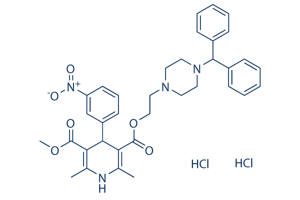
Chemische Struktur
Molekulargewicht: 683.62
Qualitätskontrolle (Quality Control)
| Verwandte Ziele | CFTR CRM1 CD markers AChR Sodium Channel Potassium Channel GABA Receptor TRP Channel ATPase GluR |
|---|---|
| Weitere Calcium Channel Inhibitoren | Bay K 8644 BAPTA-AM Tetrandrine Flunarizine 2HCl Nilvadipine Cilnidipine YM-58483 (BTP2) Ionomycin Imperatorin Astragaloside A |
Chemische Informationen, Lagerung & Stabilität (Chemical Information, Storage & Stability)
| Molekulargewicht | 683.62 | Formel | C35H38N4O6.2HCl |
Lagerung (Ab dem Eingangsdatum) | |
|---|---|---|---|---|---|
| CAS-Nr. | 89226-75-5 | SDF herunterladen | Lagerung von Stammlösungen |
|
|
| Synonyme | CV-4093 | Smiles | CC1=C(C(C(=C(N1)C)C(=O)OCCN2CCN(CC2)C(C3=CC=CC=C3)C4=CC=CC=C4)C5=CC(=CC=C5)[N+](=O)[O-])C(=O)OC.Cl.Cl | ||
Löslichkeit (Solubility)
|
In vitro |
DMSO
: 100 mg/mL
(146.28 mM)
Water : Insoluble Ethanol : Insoluble |
Molaritätsrechner
|
In vivo |
|||||
In-vivo-Formulierungsrechner (Klare Lösung)
Schritt 1: Geben Sie die untenstehenden Informationen ein (Empfohlen: Ein zusätzliches Tier zur Berücksichtigung von Verlusten während des Experiments)
Schritt 2: Geben Sie die In-vivo-Formulierung ein (Dies ist nur der Rechner, keine Formulierung. Bitte kontaktieren Sie uns zuerst, wenn es im Abschnitt "Löslichkeit" keine In-vivo-Formulierung gibt.)
Berechnungsergebnisse:
Arbeitskonzentration: mg/ml;
Methode zur Herstellung der DMSO-Stammlösung: mg Wirkstoff vorgelöst in μL DMSO ( Konzentration der Stammlösung mg/mL, Bitte kontaktieren Sie uns zuerst, wenn die Konzentration die DMSO-Löslichkeit der Wirkstoffcharge überschreitet. )
Methode zur Herstellung der In-vivo-Formulierung: Nehmen Sie μL DMSO Stammlösung, dann hinzufügenμL PEG300, mischen und klären, dann hinzufügenμL Tween 80, mischen und klären, dann hinzufügen μL ddH2O, mischen und klären.
Methode zur Herstellung der In-vivo-Formulierung: Nehmen Sie μL DMSO Stammlösung, dann hinzufügen μL Maisöl, mischen und klären.
Hinweis: 1. Bitte stellen Sie sicher, dass die Flüssigkeit klar ist, bevor Sie das nächste Lösungsmittel hinzufügen.
2. Achten Sie darauf, das/die Lösungsmittel der Reihe nach hinzuzufügen. Sie müssen sicherstellen, dass die bei der vorherigen Zugabe erhaltene Lösung eine klare Lösung ist, bevor Sie mit der Zugabe des nächsten Lösungsmittels fortfahren. Physikalische Methoden wie Vortex, Ultraschall oder ein heißes Wasserbad können zur Unterstützung des Lösens verwendet werden.
Wirkmechanismus (Mechanism of Action)
| Targets/IC50/Ki |
Calcium channel
(Guinea-pig single ventricular cells ) 2.6 nM
|
|---|---|
| In vitro |
At a holding potential of −37 mV, Manidipine decreases the Ca2+ current at concentrations above 0.1 nM, and abolishes it at 100 nM. Manidipine concentration-dependently inhibits the calcium concentration response curves. Manidipine inhibits coronay artery and renal artery with pIC50 of 9.3 nM and 9.1 nM, respectively. Manidipine partly inhibits sympathetic nerve activity and suppresses the mean arterial pressure response to infused norepinephrine. Manidipine also inhibits aldosterone secretion. Manidipine increases in both urinary calcium and uric acid. In addition, Manidipine, at nanomolar concentrations, is efficacious in modulating gene transcriptions that are involved in proinflammatory changes of mesangial cells.
|
| In vivo |
Manidipine (3 mg/kg and 10 mg/kg, p.o.) dose-dependently decreases systolic blood pressure in the three types of hypertensive rats. At the dose of 10 mg/kg, Manidipine decreases the blood pressure to the normotensive level between 1 hour and 3 hours after Manidipine is administered; the antihypertensive effect lasted for at least 8 hours. When Manidipine is administered at 10 μg/kg, the hypotensive action is markedly augmented. Manidipine potently inhibits the Ca inward current, When the potential is at -60 mV. Manidipine consistently inhibits the Ca current evoked by depolarization to 0 mV. However, a low concentration of Manidipine (1-3 nM) enhances the Ca current evoked by the depolarizing pulse of -20 mV, when the membrane potential is held at -80 mV. Inhibition of the Ca current induced by Manidipine develops slowly and over 10 minutes is required to reach the maximum inhibition.
|
Literatur |
|

Produkte sind nur für Forschungszwecke bestimmt. Nicht für den menschlichen Gebrauch. Wir verkaufen nicht an Patienten.
©Copyright 2013 Selleck Chemicals. Alle Rechte vorbehalten.