
nur für Forschungszwecke
TPCA-1 IKK-2-Inhibitor
Kat.-Nr.S2824
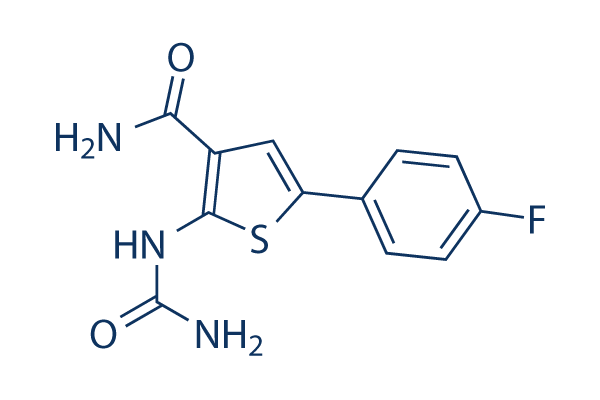
Chemische Struktur
Molekulargewicht: 279.29
Qualitätskontrolle
| Verwandte Ziele | NF-κB HDAC Antioxidant ROS Nrf2 AP-1 MALT NOD |
|---|---|
| Weitere IκB/IKK Inhibitoren | TBK1/IKKε-IN-5 Wedelolactone IKK-16 BMS-345541 Bay 11-7085 IMD 0354 MRT67307 HCl SC-514 WS6 Mesalazine (5-ASA) |
Zellkultur, Behandlung & Arbeitskonzentration
| Zelllinien | Assay-Typ | Konzentration | Inkubationszeit | Formulierung | Aktivitätsbeschreibung | PMID |
|---|---|---|---|---|---|---|
| C57BL/6 mouse BMDM cells | Cytotoxicity assay | 24 h | Cytotoxicity against C57BL/6 mouse BMDM cells assessed as LDH release after 24 hrs | 22533790 | ||
| C57BL/6 mouse BMDM cells | Function assay | 0.5 μM | 1 h | Antiinflammatory activity in C57BL/6 mouse BMDM cells assessed as inhibition of LPS-stimulated TNFalpha production at 0.5 uM pretreated for 1 hr before LPS challenge after 8 to 24 hrs by immunoassay | 22533790 | |
| MDA-MB-231 | Cytotoxicity assay | 3 days | Cytotoxicity against human MDA-MB-231 cells assessed as cell growth inhibition after 3 days by presto blue dye based plate reader method | 27077228 | ||
| TC32 | qHTS assay | qHTS of pediatric cancer cell lines to identify multiple opportunities for drug repurposing: Primary screen for TC32 cells | 29435139 | |||
| DAOY | qHTS assay | qHTS of pediatric cancer cell lines to identify multiple opportunities for drug repurposing: Primary screen for DAOY cells | 29435139 | |||
| SJ-GBM2 | qHTS assay | qHTS of pediatric cancer cell lines to identify multiple opportunities for drug repurposing: Primary screen for SJ-GBM2 cells | 29435139 | |||
| A673 | qHTS assay | qHTS of pediatric cancer cell lines to identify multiple opportunities for drug repurposing: Primary screen for A673 cells | 29435139 | |||
| SK-N-MC | qHTS assay | qHTS of pediatric cancer cell lines to identify multiple opportunities for drug repurposing: Primary screen for SK-N-MC cells | 29435139 | |||
| BT-37 | qHTS assay | qHTS of pediatric cancer cell lines to identify multiple opportunities for drug repurposing: Primary screen for BT-37 cells | 29435139 | |||
| NB-EBc1 | qHTS assay | qHTS of pediatric cancer cell lines to identify multiple opportunities for drug repurposing: Primary screen for NB-EBc1 cells | 29435139 | |||
| U-2 OS | qHTS assay | qHTS of pediatric cancer cell lines to identify multiple opportunities for drug repurposing: Primary screen for U-2 OS cells | 29435139 | |||
| Saos-2 | qHTS assay | qHTS of pediatric cancer cell lines to identify multiple opportunities for drug repurposing: Primary screen for Saos-2 cells | 29435139 | |||
| SK-N-SH | qHTS assay | qHTS of pediatric cancer cell lines to identify multiple opportunities for drug repurposing: Primary screen for SK-N-SH cells | 29435139 | |||
| NB1643 | qHTS assay | qHTS of pediatric cancer cell lines to identify multiple opportunities for drug repurposing: Primary screen for NB1643 cells | 29435139 | |||
| LAN-5 | qHTS assay | qHTS of pediatric cancer cell lines to identify multiple opportunities for drug repurposing: Primary screen for LAN-5 cells | 29435139 | |||
| Rh18 | qHTS assay | qHTS of pediatric cancer cell lines to identify multiple opportunities for drug repurposing: Primary screen for Rh18 cells | 29435139 | |||
| OHS-50 | qHTS assay | qHTS of pediatric cancer cell lines to identify multiple opportunities for drug repurposing: Primary screen for OHS-50 cells | 29435139 | |||
| RD | qHTS assay | qHTS of pediatric cancer cell lines to identify multiple opportunities for drug repurposing: Primary screen for RD cells | 29435139 | |||
| MG 63 (6-TG R) | qHTS assay | qHTS of pediatric cancer cell lines to identify multiple opportunities for drug repurposing: Primary screen for MG 63 (6-TG R) cells | 29435139 | |||
| Rh30 | qHTS assay | qHTS of pediatric cancer cell lines to identify multiple opportunities for drug repurposing: Primary screen for Rh30 cells | 29435139 | |||
| Rh41 | qHTS assay | qHTS of pediatric cancer cell lines to identify multiple opportunities for drug repurposing: Primary screen for Rh41 cells | 29435139 | |||
| A673 | qHTS assay | qHTS of pediatric cancer cell lines to identify multiple opportunities for drug repurposing: Confirmatory screen for A673 cells) | 29435139 | |||
| SK-N-MC | qHTS assay | qHTS of pediatric cancer cell lines to identify multiple opportunities for drug repurposing: Confirmatory screen for SK-N-MC cells | 29435139 | |||
| TC32 | qHTS assay | qHTS of pediatric cancer cell lines to identify multiple opportunities for drug repurposing: Confirmatory screen for TC32 cells | 29435139 | |||
| MG 63 (6-TG R) | qHTS assay | qHTS of pediatric cancer cell lines to identify multiple opportunities for drug repurposing: Confirmatory screen for MG 63 (6-TG R) cells | 29435139 | |||
| U-2 OS | qHTS assay | qHTS of pediatric cancer cell lines to identify multiple opportunities for drug repurposing: Confirmatory screen for U-2 OS cells | 29435139 | |||
| SK-N-SH | qHTS assay | qHTS of pediatric cancer cell lines to identify multiple opportunities for drug repurposing: Confirmatory screen for SK-N-SH cells | 29435139 | |||
| Klicken Sie hier, um weitere experimentelle Daten zu Zelllinien anzuzeigen | ||||||
Chemische Informationen, Lagerung & Stabilität
| Molekulargewicht | 279.29 | Formel | C12H10FN3O2S |
Lagerung (Ab dem Eingangsdatum) | |
|---|---|---|---|---|---|
| CAS-Nr. | 507475-17-4 | SDF herunterladen | Lagerung von Stammlösungen |
|
|
| Synonyme | GW683965 | Smiles | C1=CC(=CC=C1C2=CC(=C(S2)NC(=O)N)C(=O)N)F | ||
Löslichkeit
|
In vitro |
DMSO
: 56 mg/mL
(200.5 mM)
Water : Insoluble Ethanol : Insoluble |
Molaritätsrechner
|
In vivo |
|||||
In-vivo-Formulierungsrechner (Klare Lösung)
Schritt 1: Geben Sie die untenstehenden Informationen ein (Empfohlen: Ein zusätzliches Tier zur Berücksichtigung von Verlusten während des Experiments)
Schritt 2: Geben Sie die In-vivo-Formulierung ein (Dies ist nur der Rechner, keine Formulierung. Bitte kontaktieren Sie uns zuerst, wenn es im Abschnitt "Löslichkeit" keine In-vivo-Formulierung gibt.)
Berechnungsergebnisse:
Arbeitskonzentration: mg/ml;
Methode zur Herstellung der DMSO-Stammlösung: mg Wirkstoff vorgelöst in μL DMSO ( Konzentration der Stammlösung mg/mL, Bitte kontaktieren Sie uns zuerst, wenn die Konzentration die DMSO-Löslichkeit der Wirkstoffcharge überschreitet. )
Methode zur Herstellung der In-vivo-Formulierung: Nehmen Sie μL DMSO Stammlösung, dann hinzufügenμL PEG300, mischen und klären, dann hinzufügenμL Tween 80, mischen und klären, dann hinzufügen μL ddH2O, mischen und klären.
Methode zur Herstellung der In-vivo-Formulierung: Nehmen Sie μL DMSO Stammlösung, dann hinzufügen μL Maisöl, mischen und klären.
Hinweis: 1. Bitte stellen Sie sicher, dass die Flüssigkeit klar ist, bevor Sie das nächste Lösungsmittel hinzufügen.
2. Achten Sie darauf, das/die Lösungsmittel der Reihe nach hinzuzufügen. Sie müssen sicherstellen, dass die bei der vorherigen Zugabe erhaltene Lösung eine klare Lösung ist, bevor Sie mit der Zugabe des nächsten Lösungsmittels fortfahren. Physikalische Methoden wie Vortex, Ultraschall oder ein heißes Wasserbad können zur Unterstützung des Lösens verwendet werden.
Wirkmechanismus
| Targets/IC50/Ki |
NF-κB
STAT3
IKK2
(Cell-free assay) 17.9 nM
|
|---|---|
| In vitro |
In einem zeitaufgelösten Fluoreszenzresonanzenergietransfer-Assay hemmt TPCA-1 die Aktivität der humanen IKK-2 mit einer IC50 von 17,9 nM. Darüber hinaus wurde gezeigt, dass diese Verbindung ATP-kompetitiv ist. Außerdem weist sie IC50-Werte von 400 nM und 3600 nM gegen IKK-1 bzw. JNK3 auf. Diese Chemikalie hemmt die Produktion von TNF-α, IL-6 und IL-8 in einer konzentrationsabhängigen Weise, wobei IC50-Werte von 170, 290 und 320 nM erzielt werden. Sie hemmt die Proliferation von Gliomzellen sowie die TNF-induzierte nukleäre Translokation von RelA (p65) und die NFκB-abhängige IL8-Genexpression. Wichtig ist, dass diese Verbindung die IFN-induzierte Genexpression hemmt, die MX1- und GBP1-Genexpression vollständig unterdrückt, während sie nur eine geringfügige Wirkung auf die ISG15-Expression hat. |
| Kinase-Assay |
IKK-2-Assay
|
|
Rekombinantes humanes IKK-2 (Reste 1-756) wird in Baculovirus als N-terminales GST-markiertes Fusionsprotein exprimiert, und seine Aktivität wird mittels eines zeitaufgelösten Fluoreszenzresonanzenergietransfer-Assays bewertet. Kurz gesagt, IKK-2 (5 nM Endkonzentration), verdünnt in Assay-Puffer (50 mM HEPES, 10 mM MgCl2, 1 mM CHAPS, pH 7,4, mit 1 mM DTT und 0,01 % w/v BSA), wird zu Vertiefungen gegeben, die verschiedene Konzentrationen dieser Verbindung oder DMSO-Lösungsmittel (3 % Endkonzentration) enthalten. Die Reaktion wird durch Zugabe von GST-IκBα-Substrat (25 nM Endkonzentration)/ATP (1 μM Endkonzentration) in einem Gesamtvolumen von 30 μL gestartet. Die Reaktion wird 30 min bei Raumtemperatur inkubiert und dann durch Zugabe von 15 μL 50 mM EDTA beendet. Ein Detektionsreagenz (15 μL) in Puffer (100 mM HEPES, pH 7,4, 150 mM NaCl und 0,1 % w/v BSA), das den mit W-1024 Europiumchelat markierten antiphosphoserine-IκBα-32/36 monoklonalen Antikörper 12C2 und einen Allophycocyanin-markierten Anti-GST-Antikörper enthält, wird hinzugefügt, und die Reaktion wird weitere 60 min bei Raumtemperatur inkubiert. Der Phosphorylierungsgrad von GST-IκBα wird als Verhältnis des spezifischen 665-nm-Energietransfersignals zum Referenz-Europium-620-nm-Signal unter Verwendung eines Packard Discovery-Plattenlesers gemessen.
|
|
| In vivo |
Die prophylaktische Verabreichung von TPCA-1 in Dosen von 3, 10 oder 20 mg/kg, i.p., zweimal täglich, führt zu einer dosisabhängigen Verringerung der Schwere der murinen kollageninduzierten Arthritis (CIA). Die signifikant reduzierte Krankheitsaktivität und die Verzögerung des Krankheitsausbruchs, die durch die Verabreichung dieser Verbindung in einer Dosis von 10 mg/kg, i.p., zweimal täglich, erzielt werden, sind vergleichbar mit den Wirkungen des antirheumatischen Arzneimittels, i.p., jeden zweiten Tag. Die nukleäre Lokalisation von p65 sowie die Spiegel von IL-1beta, IL-6, TNF-alpha und Interferon-gamma sind im Pfotengewebe von mit dieser Verbindung behandelten Mäusen signifikant reduziert. Darüber hinaus führt die In-vivo-Verabreichung dieser Chemikalie zu einer signifikant verringerten kollageninduzierten T-Zellproliferation ex vivo. Die therapeutische Verabreichung dieser Verbindung in einer Dosis von 20 mg/kg, jedoch nicht bei 3 oder 10 mg/kg, i.p., zweimal täglich, reduziert die Schwere der CIA signifikant. |
Literatur |
|
Technischer Support
Tel: +1-832-582-8158 Ext:3
Wenn Sie weitere Fragen haben, hinterlassen Sie bitte eine Nachricht.

Produkte sind nur für Forschungszwecke bestimmt. Nicht für den menschlichen Gebrauch. Wir verkaufen nicht an Patienten.
©Copyright 2013 Selleck Chemicals. Alle Rechte vorbehalten.