
nur für Forschungszwecke
Cidofovir DNA/RNA Synthesis Inhibitor
Kat.-Nr.S1516
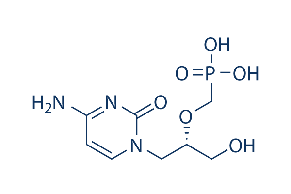
Chemische Struktur
Molekulargewicht: 279.19
Qualitätskontrolle
| Verwandte Ziele | HDAC PARP ATM/ATR DNA-PK WRN Topoisomerase PPAR Sirtuin Casein Kinase eIF |
|---|---|
| Weitere DNA/RNA Synthesis Inhibitoren | CX-5461 (Pidnarulex) B02 SCR7 Favipiravir (T-705) EED226 RK-33 BMH-21 Triapine (3-AP) Carmofur YK-4-279 |
Zellkultur, Behandlung & Arbeitskonzentration
| Zelllinien | Assay-Typ | Konzentration | Inkubationszeit | Formulierung | Aktivitätsbeschreibung | PMID |
|---|---|---|---|---|---|---|
| HEL cells | Function assay | Compound was tested for anti-viral activity against HSV-1(KOS) in HEL cells, EC50=0.57 μM | ||||
| MRC-5 cells | Function assay | Inhibitory activity against cytopathic effect of HSV-2(E 194) in MRC-5 cells, IC50=10 μM | ||||
| NHDF cells | Function assay | Inhibitory activity against replication of HCMV in NHDF cells, IC50=0.5 μM | ||||
| HFF cells | Function assay | Antiviral activity against Vaccinia virus Copenhagen in HFF cells assessed as reduction in cytopathogenicity, EC50=3.2 μM | ||||
| HFF cells | Function assay | Antiviral activity against Cowpox virus Brighton in HFF cells assessed as reduction in cytopathogenicity, EC50=7.1 μM | ||||
| HFF cells | Function assay | Antiviral activity against vaccinia virus Copenhagen measured as cytopathogenicity in HFF cells, EC50=6.9 μM | ||||
| bone marrow cells | Cytotoxicity assay | Cytotoxicity against human bone marrow cells assessed as inhibition of colony forming unit of granulocyte/macrophage, IC50=10 μM | ||||
| HFF cells | Function assay | Antiviral activity against Human CMV T2241 in HFF cells by SEAP assay, IC50=0.3 μM | ||||
| MRC5 cells | Function assay | Antiviral activity against Human CMV Towne in MRC5 cells by PRA, IC50=0.3 μM | ||||
| UC1B cells | Function assay | Antiviral activity against Murine polyomavirus MN/RDE Toronto in mouse UC1B cells assessed as reduction of virus-induced cytopathogenicity, EC50=13 μM | ||||
| african green monkey Vero cells | Function assay | Antiviral activity against Herpes simplex virus 1 F infected in african green monkey Vero cells assessed as plaque reduction after 36 to 48 hrs, EC50=14.4 μM | ||||
| HEL cells | Function assay | 3 days | Antiviral activity against HCMV AD169 infected in HEL cells assessed as reduction of virus-induced cytopathogenicity after 3 days, EC50=0.41 μM | |||
| HEL cells | Function assay | 3 days | Antiviral activity against HCMV Davis infected in HEL cells assessed as reduction of virus-induced cytopathogenicity after 3 days, EC50=0.41 μM | |||
| HFF cells | Function assay | Antiviral activity against Cytomegalovirus CMV T2211 infected in HFF cells by SEAP reporter gene assay, EC50=0.22 μM | ||||
| HEL cells | Function assay | Antiviral activity against ganciclovir-resistant HCMV AD169 clone 4 infected in HEL cells assessed as inhibition of virus-induced cytopathicity, EC50=0.74 μM | ||||
| HEL cells | Function assay | Antiviral activity against HCMV Davis infected in human HEL cells assessed inhibition of virus-induced cytopathicity after 7 days postinfection, EC50=0.5 μM | ||||
| HEL cells | Function assay | Antiviral activity against HCMV AD169 infected in human HEL cells assessed inhibition of virus-induced cytopathicity after 7 days postinfection, EC50=0.3 μM | ||||
| HEL cells | Function assay | Antiviral activity against foscarnet-resistant HCMV AD169 clone C infected in HEL cells assessed as inhibition of virus-induced cytopathicity, IC50=0.045 μM | ||||
| HFF | Proliferation assay | 3 days | Antiproliferative activity against HFF after 3 days by coulter counter assay, EC50=1.9 μM | |||
| HeLaS3 cells | Function assay | 3-5 days | Antiviral activity against Vaccinia virus IHD-J ATCC VR-156 infected in HeLaS3 cells after 3 to 5 days by plaque assay, EC50=18.74 μM | |||
| HSB2 cells | Function assay | 7 days | Antiviral activity against Human herpesvirus 6 GS infected in human HSB2 cells assessed as decrease in viral DNA accumulation after 7 days, EC50=2.7 μM | |||
| A549 cells | Function assay | Antiviral activity against Human adenovirus type 11p slobitski infected in A549 cells assessed as inhibition of DNA replication by QPCR assay, EC50=16.5 μM | ||||
| HFF cells | Function assay | Antiviral activity against Cowpox virus (Brighton Red) infected in human HFF cells assessed as reduction in viral-induced cytopathic effect by neutral red uptake assay, EC50=6.7 μM | ||||
| Klicken Sie hier, um weitere experimentelle Daten zu Zelllinien anzuzeigen | ||||||
Chemische Informationen, Lagerung & Stabilität
| Molekulargewicht | 279.19 | Formel | C8H14N3O6P |
Lagerung (Ab dem Eingangsdatum) | |
|---|---|---|---|---|---|
| CAS-Nr. | 113852-37-2 | SDF herunterladen | Lagerung von Stammlösungen |
|
|
| Synonyme | HPMPC, GS 0504 | Smiles | C1=CN(C(=O)N=C1N)CC(CO)OCP(=O)(O)O | ||
Löslichkeit
|
In vitro |
Water : 3 mg/mL
DMSO
: Insoluble
Ethanol : Insoluble |
Molaritätsrechner
|
In vivo |
|||||
In-vivo-Formulierungsrechner (Klare Lösung)
Schritt 1: Geben Sie die untenstehenden Informationen ein (Empfohlen: Ein zusätzliches Tier zur Berücksichtigung von Verlusten während des Experiments)
Schritt 2: Geben Sie die In-vivo-Formulierung ein (Dies ist nur der Rechner, keine Formulierung. Bitte kontaktieren Sie uns zuerst, wenn es im Abschnitt "Löslichkeit" keine In-vivo-Formulierung gibt.)
Berechnungsergebnisse:
Arbeitskonzentration: mg/ml;
Methode zur Herstellung der DMSO-Stammlösung: mg Wirkstoff vorgelöst in μL DMSO ( Konzentration der Stammlösung mg/mL, Bitte kontaktieren Sie uns zuerst, wenn die Konzentration die DMSO-Löslichkeit der Wirkstoffcharge überschreitet. )
Methode zur Herstellung der In-vivo-Formulierung: Nehmen Sie μL DMSO Stammlösung, dann hinzufügenμL PEG300, mischen und klären, dann hinzufügenμL Tween 80, mischen und klären, dann hinzufügen μL ddH2O, mischen und klären.
Methode zur Herstellung der In-vivo-Formulierung: Nehmen Sie μL DMSO Stammlösung, dann hinzufügen μL Maisöl, mischen und klären.
Hinweis: 1. Bitte stellen Sie sicher, dass die Flüssigkeit klar ist, bevor Sie das nächste Lösungsmittel hinzufügen.
2. Achten Sie darauf, das/die Lösungsmittel der Reihe nach hinzuzufügen. Sie müssen sicherstellen, dass die bei der vorherigen Zugabe erhaltene Lösung eine klare Lösung ist, bevor Sie mit der Zugabe des nächsten Lösungsmittels fortfahren. Physikalische Methoden wie Vortex, Ultraschall oder ein heißes Wasserbad können zur Unterstützung des Lösens verwendet werden.
Wirkmechanismus
| In vitro |
Cidofovir hemmt die Infektion mit humanem Cytomegalievirus (HCMV) in kultivierten Zellen. Diese Verbindung hemmt die CMV-Plaquebildung, selbst wenn sie den Zellen 48 Stunden nach der Infektion mit einer IC50 von 0,9 µg/mL für Davis- und 1,6 µg/mL für AD-169-Stämme zugesetzt wird. Es hemmt auch die Herpes-simplex-Virusinfektion. Darüber hinaus blockiert diese Chemikalie die durch HSV-1 induzierte Zellfusion in Affennierenzellen und blockiert die Expression von HSV-l-spezifischen Proteinen und die Synthese viraler DNA. |
|---|---|
| In vivo |
Cidofovir (5 mg/kg/Tag) subkutan über 5 Tage reduziert signifikant den durchschnittlichen Virusinfektiositätstiter in Blut, Milz, Lunge und Speicheldrüse bei infizierten Meerschweinchen. Diese Verbindung reduziert signifikant die Lymphozytose und den durchschnittlichen Gewebeindex der Milz bei infizierten Tieren. . Es unterdrückt alle Manifestationen (Hautläsionen, Lähmung der Hinterbeine und Mortalität) von haarlosen Mäusen, die intrakutan mit HSV-1 oder HSV-2 infiziert sind. Das bemerkenswerteste Merkmal dieser Verbindung ist, dass eine einmalige Verabreichung der Verbindung, selbst bis zu 4 Tage nach der Infektion, einen signifikanten Schutz gegen HSV-1- oder HSV-2-Infektion verleiht. Diese Chemikalie hemmt das Wachstum des hochaggressiven Melanomtumors, der aus Maus-Melanom-B16-Zellen entsteht, die subkutan in C57B16/J-Mäuse transplantiert wurden. |
Literatur |
|
Klinische Studieninformationen
(Daten von https://clinicaltrials.gov, aktualisiert am 2024-05-22)
| NCT-Nummer | Rekrutierung | Erkrankungen | Sponsor/Kooperationspartner | Startdatum | Phasen |
|---|---|---|---|---|---|
| NCT04542252 | Completed | Drug Drug Interaction |
SymBio Pharmaceuticals |
November 9 2020 | Phase 1 |
| NCT01610765 | Withdrawn | Herpes Simplex Virus |
University of Alabama at Birmingham |
January 2016 | Phase 1|Phase 2 |
| NCT01816646 | Completed | Blood And Marrow Transplantation |
M.D. Anderson Cancer Center|Gilead Sciences |
September 2013 | Phase 1 |
| NCT00780182 | Completed | Healthy |
Chimerix|National Institutes of Health (NIH) |
October 2008 | Phase 1 |
Technischer Support
Tel: +1-832-582-8158 Ext:3
Wenn Sie weitere Fragen haben, hinterlassen Sie bitte eine Nachricht.

Produkte sind nur für Forschungszwecke bestimmt. Nicht für den menschlichen Gebrauch. Wir verkaufen nicht an Patienten.
©Copyright 2013 Selleck Chemicals. Alle Rechte vorbehalten.