nur für Forschungszwecke
Tubastatin A HDAC Inhibitor
Kat.-Nr.S8049
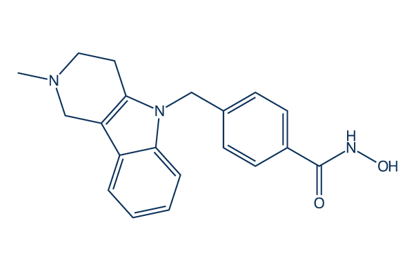
Chemische Struktur
Molekulargewicht: 335.4
Qualitätskontrolle
Zellkultur, Behandlung & Arbeitskonzentration
| Zelllinien | Assay-Typ | Konzentration | Inkubationszeit | Formulierung | Aktivitätsbeschreibung | PMID |
|---|---|---|---|---|---|---|
| Sf9 | Function assay | 2 hrs | Inhibition of full length human recombinant N-terminal GST-tagged HDAC6 (1 to 1215 residues) expressed in sf9 cells preincubated with enzyme followed by fluorogenic Arg-His-Lys-Lys(Ac)-AMC substrate addition measured after 2 hrs by fluorescence assay, IC50 = 0.0035 μM. | 27541357 | ||
| Sf9 | Function assay | Inhibition of full length human recombinant N-terminal GST-tagged HDAC6 (1 to 1215 residues) expressed in sf9 cells using RHK-K(Ac)-AMC as substrate by fluorescence assay, IC50 = 0.011 μM. | 27541357 | |||
| Sf9 | Function assay | 30 mins | Inhibition of full length human recombinant N-terminal GST-tagged HDAC6 expressed in baculovirus infected sf9 cells using fluorogenic HDAC substrate 3 after 30 mins by fluorescence assay, IC50 = 0.013 μM. | 29549837 | ||
| Sf9 | Function assay | 90 mins | Inhibition of full length human recombinant N-terminal GST-tagged HDAC6 expressed in Sf9 cells using RHKK(Ac) as substrate after 90 mins by fluorimetric method, IC50 = 0.0137 μM. | 28038324 | ||
| Sf9 | Function assay | 2 hrs | Inhibition of human recombinant HDAC6 expressed in Sf9 cells incubated for 2 hrs using RHKK-Ac fluorogenic substrate, IC50 = 0.015 μM. | 23009203 | ||
| Sf9 | Function assay | Inhibition of human recombinant HDAC6 expressed in baculovirus/sf9 cells using RHKKAc as substrate, IC50 = 0.015 μM. | 23905680 | |||
| Sf9 | Function assay | 90 mins | Inhibition of human recombinant N-terminal GST-tagged full length HDAC6 expressed in insect SF9 cells using fluorogenic ZMAL as substrate after 90 mins by fluorescence-based assay, IC50 = 0.0304 μM. | 30092367 | ||
| HeLaS3 | Function assay | 15 mins | Inhibition of HDAC6 in human HeLaS3 cells preincubated for 15 mins followed by HDAC-Glo substrate addition measured after 30 to 45 mins by ELISA, IC50 = 0.031 μM. | 28337317 | ||
| insect cells | Function assay | 4 hrs | Inhibition of human recombinant HDAC6 expressed in baculovirus infected insect cells using BATCP as substrate after 4 hrs by UHPLC-ESI-MS/MS analysis, IC50 = 0.0349 μM. | 27650925 | ||
| SHSY5Y | Function assay | 8 hrs | Inhibition of HDAC6 in human SHSY5Y cells using BATCP as substrate after 8 hrs by UHPLC-ESI-MS/MS analysis, IC50 = 0.0943 μM. | 27650925 | ||
| SHSY5Y | Function assay | 8 hrs | Inhibition of HDAC in human SHSY5Y cells using MAL as substrate after 8 hrs by UHPLC-ESI-MS/MS analysis, IC50 = 0.1221 μM. | 27650925 | ||
| Sf9 | Function assay | 4 hrs | Inhibition of full length human recombinant C-terminal FLAG/His-tagged HDAC1 expressed in baculovirus infected sf9 cells using fluorogenic HDAC substrate 3 after 4 hrs fluorescence assay, IC50 = 0.718 μM. | 29549837 | ||
| Sf9 | Function assay | Inhibition of human recombinant HDAC8 expressed in baculovirus/sf9 cells using RHKAcKAc as substrate, IC50 = 0.854 μM. | 23905680 | |||
| Sf9 | Function assay | 1 hr | Inhibition of full length human recombinant C-terminal FLAG/His-tagged HDAC1 expressed in baculovirus infected sf9 cells using fluorogenic HDAC substrate 3 after 1 hr fluorescence assay, IC50 = 0.967 μM. | 29549837 | ||
| SHSY5Y | Function assay | 8 hrs | Inhibition of HDAC1 in human SHSY5Y cells using MOCPAC as substrate after 8 hrs by UHPLC-ESI-MS/MS analysis, IC50 = 1.1097 μM. | 27650925 | ||
| Sf9 | Function assay | 30 mins | Inhibition of full length human recombinant C-terminal FLAG/His-tagged HDAC1 expressed in baculovirus infected sf9 cells using fluorogenic HDAC substrate 3 after 30 mins by fluorescence assay, IC50 = 1.54 μM. | 29549837 | ||
| Sf9 | Function assay | 90 mins | Inhibition of human recombinant C-terminal His/FLAG-tagged full length HDAC1 expressed in insect SF9 cells using fluorogenic ZMAL as substrate after 90 mins by fluorescence-based assay, IC50 = 1.91 μM. | 30092367 | ||
| HCT116 | Antiproliferative assay | 72 hrs | Antiproliferative activity against human HCT116 cells after 72 hrs by MTT assay, IC50 = 2 μM. | 27541357 | ||
| HCT116 | Antiproliferative assay | Antiproliferative activity against human HCT116 cells, IC50 = 2 μM. | 29945795 | |||
| Sf9 | Function assay | 30 mins | Inhibition of full length human recombinant C-terminal His-tagged HDAC3/N-terminal GST-tagged NCOR2 (95 to 489 residues) expressed in baculovirus infected sf9 cells using fluorogenic HDAC substrate 3 after 30 mins by fluorescence assay, IC50 = 2.26 μM. | 29549837 | ||
| HeLa | Function assay | 6 hrs | Inhibition of HDAC6 in human HeLa cells assessed as reduction in K40 hyperacetylation of alpha-tubulin incubated for 6 hrs by immunofluorescence assay, IC50 = 2.5 μM. | 25454270 | ||
| HL60 | Antiproliferative assay | 48 hrs | Antiproliferative activity against human HL60 cells after 48 hrs in presence of JAK2 inhibitor CYT-387 by CCK-8 assay, IC50 = 2.54 μM. | 29940115 | ||
| K562 | Antiproliferative assay | 48 hrs | Antiproliferative activity against human K562 cells after 48 hrs in presence of JAK2 inhibitor CYT-387 by CCK-8 assay, IC50 = 2.54 μM. | 29940115 | ||
| HEL | Antiproliferative assay | 48 hrs | Antiproliferative activity against human HEL cells after 48 hrs in presence of JAK2 inhibitor CYT-387 by CCK-8 assay, IC50 = 2.54 μM. | 29940115 | ||
| HeLaS3 | Function assay | 15 mins | Inhibition of HDAC1 in human HeLaS3 cells preincubated for 15 mins followed by HDAC-Glo substrate addition measured after 30 to 45 mins by ELISA, IC50 = 2.7 μM. | 28337317 | ||
| HeLaS3 | Function assay | 15 mins | Inhibition of HDAC3 in human HeLaS3 cells preincubated for 15 mins followed by HDAC-Glo substrate addition measured after 30 to 45 mins by ELISA, IC50 = 2.9 μM. | 28337317 | ||
| Jurkat | Cytotoxicity assay | 72 hrs | Cytotoxicity against human Jurkat cells assessed as growth inhibition after 72 hrs by MTS assay, IC50 = 3.38 μM. | 24304348 | ||
| MCF7 | Antiproliferative assay | 72 hrs | Antiproliferative activity against human MCF7 cells after 72 hrs by MTT assay, IC50 = 3.7 μM. | 27541357 | ||
| MCF7 | Antiproliferative assay | Antiproliferative activity against human MCF7 cells, IC50 = 3.7 μM. | 29945795 | |||
| HL60 | Antiproliferative assay | 48 hrs | Antiproliferative activity against human HL60 cells after 48 hrs by CCK-8 assay, IC50 = 3.75 μM. | 29940115 | ||
| K562 | Antiproliferative assay | 48 hrs | Antiproliferative activity against human K562 cells after 48 hrs by CCK-8 assay, IC50 = 3.75 μM. | 29940115 | ||
| HEL | Antiproliferative assay | 48 hrs | Antiproliferative activity against human HEL cells after 48 hrs by CCK-8 assay, IC50 = 3.75 μM. | 29940115 | ||
| HeLaS3 | Function assay | 15 mins | Inhibition of HDAC2 in human HeLaS3 cells preincubated for 15 mins followed by HDAC-Glo substrate addition measured after 30 to 45 mins by ELISA, IC50 = 3.9 μM. | 28337317 | ||
| CAL27 | Antiproliferative assay | 72 hrs | Antiproliferative activity against human CAL27 cells measured after 72 hrs by MTT assay, IC50 = 4.6 μM. | 28581289 | ||
| PC3 | Antiproliferative assay | 72 hrs | Antiproliferative activity against human PC3 cells after 72 hrs by MTT assay, IC50 = 8.6 μM. | 27541357 | ||
| PC3 | Antiproliferative assay | Antiproliferative activity against human PC3 cells, IC50 = 8.6 μM. | 29945795 | |||
| Sf9 | Function assay | 30 mins | Inhibition of full length human recombinant C-terminal His-tagged HDAC2 expressed in baculovirus infected sf9 cells using fluorogenic HDAC substrate 3 after 30 mins by fluorescence assay, IC50 = 9.97 μM. | 29549837 | ||
| MDA-MB-231 | Antiproliferative assay | 72 hrs | Antiproliferative activity against human MDA-MB-231 cells after 72 hrs by MTT assay, IC50 = 10.4 μM. | 27541357 | ||
| MDA-MB-231 | Antiproliferative assay | Antiproliferative activity against human MDA-MB-231 cells, IC50 = 10.4 μM. | 29945795 | |||
| Cal27CisR | Antiproliferative assay | 72 hrs | Antiproliferative activity against human Cal27CisR cells measured after 72 hrs by MTT assay, IC50 = 10.8 μM. | 28581289 | ||
| LNCAP | Cytotoxicity assay | 72 hrs | Cytotoxicity against androgen-dependent human LNCAP cells assessed as growth inhibition after 72 hrs by MTS assay, IC50 = 10.88 μM. | 24304348 | ||
| Cal27CisR | Function assay | 18 hrs | Inhibition of HDAC in human Cal27CisR cells using Boc-Lys(epsilon-Ac)-AMC as substrate preincubated for 18 hrs followed by substrate addition measured after 3 hrs by fluorescence assay, IC50 = 12.1 μM. | 28581289 | ||
| KB | Cytotoxicity assay | 72 hrs | Cytotoxicity against human KB cells after 72 hrs by MTS assay, IC50 = 14.81 μM. | 25899338 | ||
| THLE2 | Cytotoxicity assay | 72 hrs | Cytotoxicity against human THLE2 cells after 72 hrs by vialight cell proliferation assay, LC50 = 15.1 μM. | 29549837 | ||
| CAL27 | Function assay | 18 hrs | Inhibition of HDAC in human CAL27 cells using Boc-Lys(epsilon-Ac)-AMC as substrate preincubated for 18 hrs followed by substrate addition measured after 3 hrs by fluorescence assay, IC50 = 16.1 μM. | 28581289 | ||
| Sf9 | Function assay | 2 hrs | Inhibition of human recombinant HDAC1 expressed in Sf9 cells incubated for 2 hrs using RHKK-Ac fluorogenic substrate, IC50 = 16.4 μM. | 23009203 | ||
| Sf9 | Function assay | Inhibition of human recombinant HDAC1 expressed in baculovirus/sf9 cells using RHKKAc as substrate, IC50 = 16.4 μM. | 23905680 | |||
| B16 | Growth inhibition assay | 48 hrs | Growth inhibition of mouse B16 cells incubated for 48 hrs by MTT assay, GI50 = 40.5 μM. | 23009203 | ||
| KB | Function assay | 14 uM | 24 hrs | Inhibition of HDAC1 in human KB cells assessed as increase in histone H4 acetylation at 14 uM after 24 hrs by Western blotting analysis | 25899338 | |
| KMS-12-BM | Function assay | 15 uM | up to 48 hrs | Inhibition of HDAC6 in human KMS-12-BM cells assessed as increase in acetylated tubulin level at 15 uM up to 48 hrs by immunoblot method | 27541357 | |
| MOLM14 | Function assay | 15 uM | up to 48 hrs | Inhibition of HDAC6 in human MOLM14 cells assessed as increase in acetylated tubulin level at 15 uM up to 48 hrs by immunoblot method | 27541357 | |
| U937 | Function assay | 2 uM | 18 hrs | Inhibition of HDAC6 in human U937 cells assessed as increase in alpha-tubulin acetylation at Lys-40 residue at 2 uM after 18 hrs by Western blot method | 28337317 | |
| LNCAP | Function assay | 24 hrs | Inhibition of HDAC6 in human LNCAP cells assessed as inhibition of DHT-induced alpha-tubulin deacetylation by measuring increase in alpha-tubulin acetylation measured after 24 hrs relative to control | 27717544 | ||
| human | Function assay | 24 hrs | Antagonist activity at AR in human LNCAP cells assessed as suppression of DHT-induced AR protein level measured after 24 hrs relative to control | 27717544 | ||
| DAOY | qHTS assay | qHTS of pediatric cancer cell lines to identify multiple opportunities for drug repurposing: Primary screen for DAOY cells | 29435139 | |||
| SJ-GBM2 | qHTS assay | qHTS of pediatric cancer cell lines to identify multiple opportunities for drug repurposing: Primary screen for SJ-GBM2 cells | 29435139 | |||
| A673 | qHTS assay | qHTS of pediatric cancer cell lines to identify multiple opportunities for drug repurposing: Primary screen for A673 cells | 29435139 | |||
| SK-N-MC | qHTS assay | qHTS of pediatric cancer cell lines to identify multiple opportunities for drug repurposing: Primary screen for SK-N-MC cells | 29435139 | |||
| BT-37 | qHTS assay | qHTS of pediatric cancer cell lines to identify multiple opportunities for drug repurposing: Primary screen for BT-37 cells | 29435139 | |||
| NB-EBc1 | qHTS assay | qHTS of pediatric cancer cell lines to identify multiple opportunities for drug repurposing: Primary screen for NB-EBc1 cells | 29435139 | |||
| U-2 OS | qHTS assay | qHTS of pediatric cancer cell lines to identify multiple opportunities for drug repurposing: Primary screen for U-2 OS cells | 29435139 | |||
| Saos-2 | qHTS assay | qHTS of pediatric cancer cell lines to identify multiple opportunities for drug repurposing: Primary screen for Saos-2 cells | 29435139 | |||
| SK-N-SH | qHTS assay | qHTS of pediatric cancer cell lines to identify multiple opportunities for drug repurposing: Primary screen for SK-N-SH cells | 29435139 | |||
| NB1643 | qHTS assay | qHTS of pediatric cancer cell lines to identify multiple opportunities for drug repurposing: Primary screen for NB1643 cells | 29435139 | |||
| LAN-5 | qHTS assay | qHTS of pediatric cancer cell lines to identify multiple opportunities for drug repurposing: Primary screen for LAN-5 cells | 29435139 | |||
| Rh18 | qHTS assay | qHTS of pediatric cancer cell lines to identify multiple opportunities for drug repurposing: Primary screen for Rh18 cells | 29435139 | |||
| OHS-50 | qHTS assay | qHTS of pediatric cancer cell lines to identify multiple opportunities for drug repurposing: Primary screen for OHS-50 cells | 29435139 | |||
| RD | qHTS assay | qHTS of pediatric cancer cell lines to identify multiple opportunities for drug repurposing: Primary screen for RD cells | 29435139 | |||
| Rh30 | qHTS assay | qHTS of pediatric cancer cell lines to identify multiple opportunities for drug repurposing: Primary screen for Rh30 cells | 29435139 | |||
| Rh41 | qHTS assay | qHTS of pediatric cancer cell lines to identify multiple opportunities for drug repurposing: Primary screen for Rh41 cells | 29435139 | |||
| HEL | Cell cycle assay | 1 to 5 uM | 48 hrs | Cell cycle arrest in human HEL cells assessed as accumulation at G1 phase at 1 to 5 uM after 48 hrs propidium iodide staining based flow cytometry | 29940115 | |
| HeLa | Function assay | 2 uM | 12 hrs | Inhibition of HDAC6 in human HeLa cells assessed as increase in acetyl-tubulin level at 2 uM after 12 hrs by Western blot analysis | 29533873 | |
| HEK293 | Function assay | 10 uM | 24 hrs | Inhibition of HDAC1 in HEK293 cells assessed as increase in histone H3 acetylation at 10 uM after 24 hrs by Western blot method | 28523102 | |
| MV4-11 | Function assay | 200 nM | 24 hrs | Inhibition of HDAC6 in human MV4-11 cells assessed as accumulation of acetylated alpha-tubulin at 200 nM after 24 hrs by Western blot analysis | 29738953 | |
| HCT116 | Cell cycle assay | 5 uM | 48 hrs | Cell cycle arrest in human HCT116 cells assessed as accumulation at sub-G1 phase at 5 uM after 48 hrs by propidium iodide staining-based flow cytometric method | 28038324 | |
| PC12 | Neuroprotective assay | 10 uM | 24 hrs | Neuroprotective activity against H2O2-induced toxicity in rat PC12 cells assessed as cell viability at 10 uM pretreated for 24 hrs followed by H2O2 challenge and measured after 12 hrs by MTT assay relative to control | 30385227 | |
| PC12 | Neuroprotective assay | 5 uM | 24 hrs | Neuroprotective activity against 6-OHDA-induced toxicity in rat PC12 cells assessed as increase in cell viability at 5 uM pretreated for 24 hrs followed by 6-OHDA challenge and measured after 12 hrs by MTT assay | 30385227 | |
| PC12 | Neuroprotective assay | 10 uM | 24 hrs | Neuroprotective activity against 6-OHDA-induced toxicity in rat PC12 cells assessed as increase in cell viability at 10 uM pretreated for 24 hrs followed by 6-OHDA challenge and measured after 12 hrs by MTT assay | 30385227 | |
| PC12 | Cytoprotective assay | 24 hrs | Cytoprotective activity against H2O2-induced damage in rat PC12 cells assessed as decrease in ROS accumulation preincubated for 24 hrs followed by H2O2 challenge measured after 12 hrs by DCFH-DA dye-based fluorescence analysis | 30385227 | ||
| PC12 | Cytoprotective assay | 24 hrs | Cytoprotective activity against H2O2-induced damage in rat PC12 cells assessed as decrease in ROS accumulation preincubated for 24 hrs followed by H2O2 challenge measured after 12 hrs by DCFH-DA dye-based inverted fluorescence microscopic analysis | 30385227 | ||
| PC12 | Neuroprotective assay | 5 to 10 uM | 24 hrs | Neuroprotective activity against 6-OHDA-induced toxicity in rat PC12 cells assessed as increase in cell viability at 5 to 10 uM pretreated for 24 hrs followed by 6-OHDA challenge and measured after 12 hrs coincubated with ebselen by MTT assay | 30385227 | |
| PC12 | Antioxidant assay | 5 uM | 24 hrs | Antioxidant activity against H2O2-induced oxidative stress in rat PC12 cells assessed as decrease in ROS accumulation at 5 uM preincubated for 24 hrs followed by H2O2 challenge and measured after 12 hrs by DCFH-DA dye-based fluorescence analysis | 30385227 | |
| PC12 | Antioxidant assay | 5 uM | 24 hrs | Antioxidant activity against H2O2-induced oxidative stress in rat PC12 cells assessed as decrease in ROS accumulation at 5 uM preincubated for 24 hrs followed by H2O2 challenge and measured after 12 hrs coincubated with ebselen by DCFH-DA dye-based fluore | 30385227 | |
| Klicken Sie hier, um weitere experimentelle Daten zu Zelllinien anzuzeigen | ||||||
Chemische Informationen, Lagerung & Stabilität
| Molekulargewicht | 335.4 | Formel | C20H21N3O2 |
Lagerung (Ab dem Eingangsdatum) | |
|---|---|---|---|---|---|
| CAS-Nr. | 1252003-15-8 | SDF herunterladen | Lagerung von Stammlösungen |
|
|
| Synonyme | N/A | Smiles | CN1CCC2=C(C1)C3=CC=CC=C3N2CC4=CC=C(C=C4)C(=O)NO | ||
Löslichkeit
|
In vitro |
DMSO
: 16.7 mg/mL
(49.79 mM)
Water : Insoluble Ethanol : Insoluble |
Molaritätsrechner
|
In vivo |
|||||
In-vivo-Formulierungsrechner (Klare Lösung)
Schritt 1: Geben Sie die untenstehenden Informationen ein (Empfohlen: Ein zusätzliches Tier zur Berücksichtigung von Verlusten während des Experiments)
Schritt 2: Geben Sie die In-vivo-Formulierung ein (Dies ist nur der Rechner, keine Formulierung. Bitte kontaktieren Sie uns zuerst, wenn es im Abschnitt "Löslichkeit" keine In-vivo-Formulierung gibt.)
Berechnungsergebnisse:
Arbeitskonzentration: mg/ml;
Methode zur Herstellung der DMSO-Stammlösung: mg Wirkstoff vorgelöst in μL DMSO ( Konzentration der Stammlösung mg/mL, Bitte kontaktieren Sie uns zuerst, wenn die Konzentration die DMSO-Löslichkeit der Wirkstoffcharge überschreitet. )
Methode zur Herstellung der In-vivo-Formulierung: Nehmen Sie μL DMSO Stammlösung, dann hinzufügenμL PEG300, mischen und klären, dann hinzufügenμL Tween 80, mischen und klären, dann hinzufügen μL ddH2O, mischen und klären.
Methode zur Herstellung der In-vivo-Formulierung: Nehmen Sie μL DMSO Stammlösung, dann hinzufügen μL Maisöl, mischen und klären.
Hinweis: 1. Bitte stellen Sie sicher, dass die Flüssigkeit klar ist, bevor Sie das nächste Lösungsmittel hinzufügen.
2. Achten Sie darauf, das/die Lösungsmittel der Reihe nach hinzuzufügen. Sie müssen sicherstellen, dass die bei der vorherigen Zugabe erhaltene Lösung eine klare Lösung ist, bevor Sie mit der Zugabe des nächsten Lösungsmittels fortfahren. Physikalische Methoden wie Vortex, Ultraschall oder ein heißes Wasserbad können zur Unterstützung des Lösens verwendet werden.
Wirkmechanismus
| Targets/IC50/Ki |
HDAC6
(Cell-free assay) 15 nM
|
|---|---|
| In vitro |
Tubastatin A ist selektiv gegenüber allen Isozymen außer HDAC8 und behält eine über 1000-fache Selektivität gegenüber allen Isoformen, ausgenommen HDAC8, wo es eine ungefähr 57-fache Selektivität aufweist. Diese Verbindung induziert bevorzugt die α-Tubulin-Hyperacetylierung bei 2,5 μM. Eine leichte Induktion der Histon-Hyperacetylierung wird für diese Chemikalie bei 10 μM beobachtet. Es zeigt einen dosisabhängigen Schutz gegen Homocysteinsäure-induzierten neuronalen Zelltod, beginnend bei 5 μM mit nahezu vollständigem Schutz bei 10 μM.
Diese Verbindung (10 μM) induziert eine Zunahme der acetylierten α-Tubulin-Spiegel und die Wiederherstellung der primären Zilienexpression in den Cholangiokarzinom-Zelllinien (18-fach); und die Wiederherstellung der primären Zilien korrelierte mit herunterregulierten Hedgehog (Hh)- und MAPK-Signalwegen, sowie verringerten Zellproliferationsraten (im Durchschnitt um 50%) und Invasion (um 40%).
Es zeigt eine signifikante Hemmung von TNF-α und IL-6 in LPS-stimulierten menschlichen THP-1-Makrophagen mit einer IC50 von 272 nM und 712 nM. Dieser Inhibitor hemmt die Stickoxid (NO)-Sekretion in murinen Raw 264.7-Makrophagen dosisabhängig mit einer IC50 von 4,2 μM.
|
| Kinase-Assay |
HDAC-Enzymtests
|
|
Tubastatin A wird in Assay-Puffer (50 mM HEPES, pH 7,4, 100 mM KCl, 0,001% Tween-20, 0,05% BSA und 20 μM Tris(2-carboxyethyl)phosphin) gelöst und auf das 6-fache der Endkonzentration verdünnt. HDAC-Enzyme werden in Assay-Puffer auf das 1,5-fache der Endkonzentration verdünnt und 10 Minuten lang mit dieser Verbindung vor Zugabe des Substrats vorinkubiert. Die Menge an FTS (HDAC1, HDAC2, HDAC3 und HDAC6) oder MAZ-1675 (HDAC4, HDAC5, HDAC7, HDAC8 und HDAC9), die für jedes Enzym verwendet wird, entspricht der Michaelis-Konstante (Km), wie durch eine Titrationskurve bestimmt. FTS oder MAZ-1675 wird in Assay-Puffer auf das 6-fache der Endkonzentration mit 0,3 μM Trypsin in Sequenzierungsqualität verdünnt. Das Substrat/Trypsin-Gemisch wird dem Enzym/Verbindungs-Gemisch zugesetzt, die Platte wird 60 Sekunden lang geschüttelt und dann in einen SpectraMax M5 Mikrotiterplattenleser gestellt. Die enzymatische Reaktion wird 30 Minuten lang auf die Freisetzung von 7-Amino-4-methoxycoumarin überwacht, nach Deacetylierung der Lysinseitenkette im Peptidsubstrat, und die lineare Rate der Reaktion wird berechnet.
|
|
| In vivo |
Tubastatin A reduziert das Wachstum von Cholangiokarzinomen in vivo. Diese Verbindung (10 mg/kg) induziert ein 6-fach niedrigeres mittleres Tumorgewicht in einem syngenen orthotopen Rattenmodell für Cholangiokarzinom und eine Reduzierung der Verhältnisse von Tumorgewicht zu Lebergewicht und Körpergewicht (5- bzw. 5,6-fach) sowie eine höhere Häufigkeit von ziliierten Cholangiozyten im Vergleich zu Kontrollen (29% vs. 1,4%). Es verringert signifikant die Anzahl der PCNA-positiven Zellen in den behandelten Tumoren im Vergleich zu Vehikelkontrollen (34% vs. 65%).
Diese Chemikalie zeigt eine signifikante Hemmung des Pfotenvolumens bei 30 mg/kg i.p. in einem durch Freunds vollständiges Adjuvans (FCA) induzierten Tiermodell der Entzündung. Es (30 mg/kg i.p.) attenuisiert signifikant die klinischen Scores (~ 70%) und die IL-6-Expression in Pfotengeweben von Kollagen-induzierter Arthritis bei DBA1-Mäusen.
|
Literatur |
|
Anwendungen
| Methoden | Biomarker | Bilder | PMID |
|---|---|---|---|
| Western blot | EGFR / p-AKT / AKT / p-ERK / ERK |
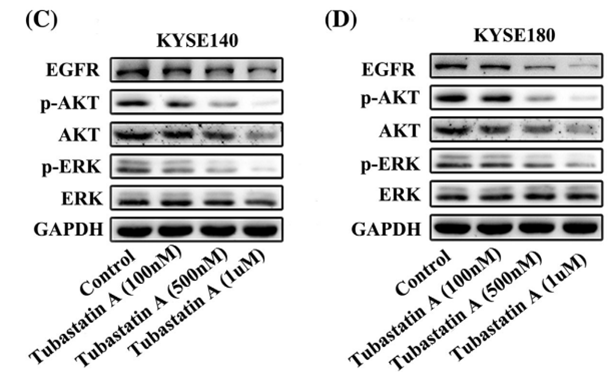
|
29665050 |
| Immunofluorescence | α-tubulin / Acetylated tubulin HDAC6 |
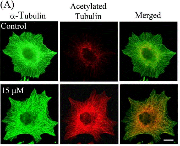
|
23798680 |
Technischer Support
Tel: +1-832-582-8158 Ext:3
Wenn Sie weitere Fragen haben, hinterlassen Sie bitte eine Nachricht.
Häufig gestellte Fragen
Frage 1:
We are planning to order some, but I found out there are two versions of it. One has HCl and one does not. Which one do you recommend for live cell use? Will the HCl containing version significantly change the pH?
Antwort:
S8049 and S2627 have the same molecular structure. The only difference is S2627 containing HCl and has higher solubility in DMSO (74 mg/mL vs. S8049 9 mg/mL). Since they are the same molecule, its biological function should be similar. I would recommend to use S2627 for cell culture study.
Frage 2:
What vehicle do you recommend to dissolve it for in vivo experiments?
Antwort:
It can be dissolved in 2% DMSO/30% PEG 300/PBS at 2.5 mg/mL as a clear solution, and is also a clear solution in 2% DMSO/ corn oil at 2.5 mg/mL. This compound in 2% DMSO/0.5% Tween 80/PBS is a homogeneous suspension at 2.5 mg/mL at first. After stay for a while, the precipitation goes out at the bottom of the tube.
Produkte sind nur für Forschungszwecke bestimmt. Nicht für den menschlichen Gebrauch. Wir verkaufen nicht an Patienten.
©Copyright 2013 Selleck Chemicals. Alle Rechte vorbehalten.